A dedicated group of geologists, physicists, archaeologists, political scientists, biologists, english and history majors joined forces to learn a bit about Climate Change in the natural laboratory of Northeast Ohio. Here they surround a glacial erratic in Secrest Arboretum of the OARDC – where The Ohio State University and the National Weather Service has meteorological records extending back to the late 1800s CE. The Arboretum also has an extensive collection of stands of trees from around the world that are used in our climate studies below (special thanks to Joe Cochran (OSU) for permission to work at Secrest).
The first project: the glacial transition in a sediment core from Browns Lake Bog

Dr. Thomas Lowell gives the rundown at Browns Lake Bog – Tom is a professor at the University of Cincinnati and long-time collaborator and the core boss.

Initial description of the 5 meter core – we obtained two radiocarbon ages, measured magnetic susceptibility, loss on ignition, in addition to core description and sediment analyses.
The Upshot of the Lake Work – The two ages were chosen at transitions in the character of the peat and mineral matter – we identified a major shift at the time of the Bolling – Allerod warming and at the cooling of the Younger Dryas. The abrupt climate changes (ACCs) and discussion of how the world moves from the Pleistocene to the Holocene is brought home to Ohio in this core (Figure below). It is exciting to explore how these ACCs affected NE-Ohio’s ecosystems and physical landscapes.

Project 2: Tree Ring Dating of the Biggio Barn

The barn owner gives the rundown on the history and possible ages of the hand hewn timber frame. The dating of the barn project introduced the class to the science of tree-rings.

Hong Kong dendrochronologist, Vincent shows the class how by standing on two milk crates he cores a beam – the instructor adds a stabilizing foot to Vincent’s precarious sampling strategy.
The upshot of Barn Dating: Ten of the beams from the Biggio Barn were cut in the spring of 1840 CE. The building then was likely constructed shortly after that cut date. A copy of the report to the owner from the class can be found here. The ring-width data obtained in this study are used in drought studies below. The Wooster Tree Ring lab has dated over 60 barns and houses in Ohio and PA (this video describes the process and some of the science).
Project 3a: Extracting a Temperature Proxy Record from Larch in Kamchatka
Vincent Hui, Abbey Martin, Sarah McGrath, Matthew Shearer, Ann Wilkinson
The purpose of this study was to analyze Kamchatka larch (Larix cajandery Mayr.) tree ring widths from Fareast, Russia. The team standardized the chronology using two methods, (1) negative exponential, and (2) regional curve standardization (RCS), and they then compared how the standardization technique influenced correlations. Both standardized series were correlated with meteorological records showing high positive correlations for summer temperatures. The RCS showed stronger correlations and was used for NTREND comparison, temperature reconstruction, and spectral analysis. Together these correlations and comparisons showed the larch primarily responds to summer temperature and can be used to reconstruct summer temperatures.

The Kamchatka team of researchers (without Vincent) who did the study. They are posing at Wooster Memorial Park where a recent planting of 700 trees and prairie will sequester more carbon in the future than the previous agricultural land use at the site.

 Conclusions:
1 – The Kamchatka larch tree-ring widths are most sensitive to summer (May through September) temperatures.
2 – The team recommends the region curve standardization method) RCS method for standardization with a sample size of 190 series.
3 – The RCS series showed similar trends as the NTREND series, suggesting the Kamchatka site follows the same trends as much of the northern hemisphere.
4 – Ring-widths show a general increase in temperature over the last 350 years for the interior of Kamchatka. This is unprecedented over the past 300 years and is consistent with other proxies such as glaciers.
Project 3b: Past climate inferences using data from Johnson Woods
Sharron Osterman, Annette Hilton, Cameron Steckbeck, Gina Malfatti, Amineh AlBashair

- The Johnson Woods team assembled a newly compiled data set originally sampled in 1985 by Dr. Ed Cook (LDEO), by the Wooster Tree Ring Lab in 2003 and most recently updated by Dr. Justin Maxwell (Indiana State University). They found there was a marked release in the tree ring record across northern Ohio about the time of European Settlement in the region. This may be in part due to the disturbance in the record, however it could also persist due to the positive response that tree growth has to summer precipitation.

 Above is a histogram showing the correlations of the Johnson Woods ring-width series and monthly precipitation and temperature records from the OARDC spanning 1880 to 2014 CE. The trees are a record of summer precipitation (positive correlation) and favor wet summers. These trees are negatively correlated with high summer temperatures.
One Question on the final exam:
What is the Climate response of European Larch to climate of Ohio – Secrest Arboretum (and why might this exploration be relevant?).
Obtaining high quality cores for ring-width chronologies from European Larch at Secrest Arboretum.

The upshot here is the ring-width chronology below. The class worked on this as part of the final exam and found that similar to the oaks in the region, the European Larch is sensitive to summer precipitation and is stressed by high summer temperatures. The tailing off of the ring-widths during recent decades could be the result of warmer summer temperatures – a hypothesis that needs testing. The relevance of this study is that as climate changes in the high latitudes of Europe and Asia, where these larch dominate – it may be the case, that warming may stress the species leading to decreases in bioproductivity – these ideas need further work to test if this is a viable hypothesis.


A day in Johnson Woods – the full class in the rain.

 We also learned that Dan Misinay (’16) is a pretty fair teaching assistant.

The class wanders around the gas power plant on the Wooster campus – three years ago the college transitioned from coal burning to natural gas – the carbon dioxide emissions on campus have been cut in half. However, now the College buys its power for cooling (air conditioning) off campus from the grid, where much of the electricity is powered by coal, but with a growing portfolio of clean energy sources (special thanks to Lanny Whitaker who showed us the plant and explained where our energy comes from – thank you). We also thank Nick Wiesenberg (our able Geology Technician) for his knowledge of trees, barn dating and general troubleshooting, Tom Lowell and his students for the high quality sediment cores, our TA Dan and a host of tree-ring scientists who contributed data to our efforts in this course. Special thanks too – to the Secrest Arboretum. A portion of the Kamchatka tree-ring record was supported by NSF- AGS – 1202218.


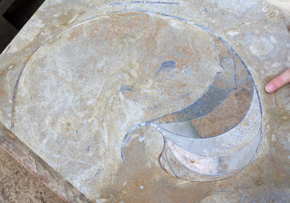
 This past semester I worked with three colleagues on a massive trace fossil review paper, which we hope meets success in the next month or so. My primary job on the team was to sort out bioerosion traces, especially those that are macroscopic. As always with such studies, I learned a great deal when forced to do a systematic literature review. One of the ichnogenera new to me was Caedichnus, a wedge-shaped excision found primarily in gastropod shells. It was only described last year by Stafford et al. (2015). Above is an example we happened to have in our collections. Note the fractured margins in this Fusinus shell aperture from the Pliocene of Cyprus. It was likely made by a predatory crustacean (such as a crab or lobster) bashing away at the shell to get to the living snail inside. The predator may have been successful in this case since there is no sign of healing in the snail shell.
 Above is an undamaged Fusinus showing a complete aperture. This snail also had its travails, though. Note the round, incomplete borehole just above the aperture. This was made by some kind of drilling predator, likely a naticid snail.