Wooster’s Pseudofossils of the Week: Shatter cones from southern Ohio

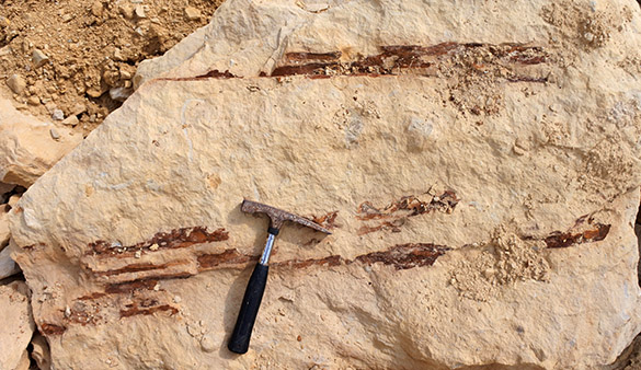
Real shatter cones 585This brief post is a correction of a previous entry. Last year I showed what I thought were shatter cones collected many years ago in Adams County, Ohio, by the late Professor Frank L. Koucky of The College of Wooster. James Chesire commented on the post and said it was more likely the specimens were cone-in-cone structures produced by burial diagenesis not bolide impacts. When he sent me the photo above of real shatter cones from the Serpent Mound impact region in southern Ohio, I knew he was correct. Shatter cones have distinctive radiating, longitudinal fractures not seen in similar conical structures in limestones. The above shatter cones are in an unknown Ordovician limestone.

Both shatter cones and cone-in-cone structures are nevertheless pseudofossils in that they are both sometimes confused with organic structures like corals and chaetetids. I shall never mix them up again! Thanks for the correction, James.

References:

Carlton, R.W., Koeberl, C., Baranoski, M.T. and Schumacher, G.A. 1998. Discovery of microscopic evidence for shock metamorphism at the Serpent Mound structure, south-central Ohio: confirmation of an origin by impact. Earth and Planetary Science Letters 162: 177-185.

Dietz, R.S. 1959. Shatter cones in cryptoexplosion structures (meteorite impact?). The Journal of Geology 67: 496-505.

Sagy, A., Fineberg, J. and Reches, Z. 2004. Shatter cones: Branched, rapid fractures formed by shock impact. Journal of Geophysical Research 109: B10209.

Shaub, B.M. 1937. The origin of cone-in-cone and its bearing on the origin of concretions and septaria. American Journal of Science 203: 331-344.

Posted in Uncategorized | Tagged , , , | 1 Comment

Wooster’s Fossil of the Week: A low-spired, battle-worn trochid gastropod from the Pliocene of Cyprus

1 Gibbula Risso, 1826 apexThis shell looks like a cinnamon roll. It is another product of the 1996 Wooster-Keck expedition to Cyprus with Steve Dornbos (’97) and me. Like the rest of the Cypriot specimens on this blog, it is from the Nicosia Formation (Pliocene) exposed on the Mesaoria Plain in the center of the island. This specimen comes from the Coral Reef locality described in Dornbos and Wilson (1999). We are looking at a well-preserved specimen of the herbivorous gastropod Gibbula Risso, 1826. (I can’t fit it into any known species within the genus.)
2 Gibbula Risso, 1826 sideIn this side view the growth lines are evident (they are parallel to the aperture; the thin ribs following the whorls are ornamentation), as are a couple of shallow, circular pits drilled by some unsuccessful predator. That predator could have been another gastropod or even an octopus. The pits are known by the trace fossil name Oichnus.

Those growth lines are interesting in  this genus. Schöne et al. (2007) studied a species of modern Gibbula and determined that they formed “microgrowth lines” in association with tidal cycles, forming “distinct fortnight bundles of microgrowth increments and lines”. We would need to section this shell and examine it microscopically to see such patterns.

3 Gibbula Risso, 1826 baseHere is the basal view of our Gibbula specimen.

4 RissoThe genus Gibbula was named and described by Giuseppe Antonio Risso (1777-1845), called Antoine Risso, was a productive Italian (more or less; he later can be considered French) naturalist. He was born in the city of Nice, then in the Duchy of Savoy. In 1792, soon after the French Army occupied Nice, Risso became a pharmacist’s apprentice, which encouraged his interest in medicinal botany. Risso was also a pioneering mountaineer in the Alps and other European ranges. He published several books on invertebrates, fish and plants. The work most relevant to us is his 1826 tome entitled: Histoire naturelle des principales productions de l’Europe Méridionale et particulièrement de celles des environs de Nice et des Alpes Maritimes. Risso’s Dolphin is named after him.

References:

Donnarumma, L., Bruno, R., Terlizzi, A. and Russo, G.F. 2015. Population ecology of Gibbula umbilicaris and Gibbula ardens (Gastropoda: Trochidae) in a Posidonia oceanica seagrass bed. Italian Journal of Zoology, DOI: 10.1080/11250003.2015.1073377

Dornbos, S.Q. and Wilson, M.A. 1999. Paleoecology of a Pliocene coral reef in Cyprus: Recovery of a marine community from the Messinian Salinity Crisis. Neues Jahrbuch für Geologie und Paläontologie, Abhandlungen 213: 103-118.

Risso, A. 1826. Histoire naturelle des principales productions de l’Europe Méridionale et particulièrement de celles des environs de Nice et des Alpes Maritimes. Paris: F.G. Levrault. Vol. 4: IV, 1-439, 12 pls.

Schöne, B.R., Rodland, D.L., Wehrmann, A., Heidel, B., Oschmann, W., Zhang, Z., Fiebig, J. and Beck, L. 2007. Combined sclerochronologic and oxygen isotope analysis of gastropod shells (Gibbula cineraria, North Sea): life-history traits and utility as a high-resolution environmental archive for kelp forests. Marine Biology 150: 1237-1252.

Williams, E.E. 1964. The growth and distribution of Gibbula umbilicalis (da Costa) on a rocky shore in Wales. The Journal of Animal Ecology 33: 433-442.

Posted in Uncategorized | Tagged , , , | Leave a comment

Wooster’s Fossil of the Week: A conid gastropod from the Pliocene of Cyprus

Conus pelagicus Epsilos 585Cyprus again for this week’s fossil. This is a nearly complete shell of the predatory snail Conus pelagicus Brocchi 1814 found at the Epsilos exposure of the Nicosia Formation (Pliocene) on the Mesaoria Plain of central Cyprus by Steve Dornbos (’97) and me in 1996. In life this species no doubt had an intricate shell color pattern, as their cousins do today.

The taxonomic intricacies of the genus Conus are far beyond the scope of a mere blog entry, so I’ll simply link to a list of associated genera, subgenera and synonymies. Conus as an organism is fantastic. These are venomous predators famous for shooting radular teeth loaded with very effective toxins. Some species can kill a human in less than five minutes. No worries, though — the venom contains analgesic compounds so there is little pain. The best way to demonstrate the extraordinary killing process used by Conus is to look at a video. You’ll never look at snails the same way again.
BrocchiImageConus pelagicus was originally described by Giovanni Battista Brocchi in 1814. We met him in a previous blog entry, so much of this information is repeated. Brocchi (1772-1826) was an Italian natural historian who made significant contributions to botany, paleontology, mineralogy and general geology. He was born in Bassano del Grappa, Italy, and studied law at the University of Padova. He liked mineralogy and plants much better than lawyering, though, and became a professor in Brescia. His work resulted in an appointment as Inspector of Mines in the new kingdom of Italy. He famously said, “The science of fossil shells is the first step towards the study of the earth.”

Brocchi wrote the first thorough geological assessment of the Apennine Mountains, and he included in it a remarkable systematic study of Neogene fossils. He compared these fossils to modern animals in the Mediterranean — a very progressive thing to do at the time.
Brocchi plate 122915Above are drawings made by Brocchi of the conid (and a couple cypraeid) fossils he found in the Apennines during his extensive study published in 1814. Note that in the Continental fashion still followed today, the shells are figured aperture-up. Americans and the rest of the English-speaking world orient them in the proper way. Figures 11a, b and c, though, are oriented in the opposite direction, maybe to fill the space efficiently.

Brocchi was an adventurous traveler, but it eventually did him in. He died in Khartoum in 1826, a “victim of the climate” and a martyr for field science.

References:

Brocchi, G.B. 1814. Conchiologia fossile subapennina con osservazioni geologiche sugli Apennini e sul suolo adiacente. Milano Vol. I: pp. LXXX + 56 + 240; Vol. II, p. 241-712, pl. 1-16.

Cowper Reed, F.R. 1935. Notes on the Neogene faunas of Cyprus, III: the Pliocene faunas. Annual Magazine of Natural History 10 (95): 489-524.

Cowper Reed, F.R. 1940. Some additional Pliocene fossils from Cyprus. Annual Magazine of Natural History 11 (6): 293-297.

Dornbos, S.Q. and Wilson, M.A. 1999. Paleoecology of a Pliocene coral reef in Cyprus: Recovery of a marine community from the Messinian Salinity Crisis. Neues Jahrbuch für Geologie und Paläontologie, Abhandlungen 213: 103-118.

Posted in Uncategorized | Tagged , , , | 5 Comments

Wooster’s Fossil of the Week: A muricid gastropod from the Pliocene of Cyprus

1 Bolinus brandaris coral reef 585We return to Cyprus for this week’s fossil. This is a broken shell of the predatory muricid Bolinus brandaris (Linnaeus, 1758) found at the Coral Reef exposure of the Nicosia Formation (Pliocene) on the Mesaoria Plain of central Cyprus by Steve Dornbos (’97) and myself in 1996. It has had some significant battering, probably by a crab peeling away the shell to get to the goodies. The ribs are prominent on the whorls, representing previous strengthened apertures. The base was a long, narrow siphonal canal now broken off. You can see the complete shell shape in modern examples.
2 Bolinus brandaris opened coral reef 585On the other side of the same specimen we see that the last whorl has been opened, showing the spiral columella at the axis of coiling. The arrow points to a puncture hole in an upper whorl. This appears to be the result of a “ballistic” impact on the shell by some predatory organism. Pether (1995) gave the name Belichnus to this trace fossil, attributing it to stomatopod crustaceans and their wicked-fast and powerful claws. Cadée and de Wolf (2013) expanded the possible tracemakers of Belichnus to seagulls.

Bolinus brandaris is well known today throughout the Mediterranean as the Purple Dye Murex. It has been used since ancient times as the source of a deep, permanent fabric dye called Tyrian Purple. It was highly prized by royalty and the wealthy elite for millennia.

Linnaeus originally placed this species in the genus Murex, but in 1837 Georg Gottlieb Pusch described a new but similar genus Bolinus, to which the species now belongs. Pusch, who also had the Polish name Jerzy Bogumił Koreński, was a very interesting fellow in the early days of paleontology and geology. He was born in Kohren, Saxony, in 1790 (or 1791, depending on which calendar you use). He was very early interested in what would become geology, so in 1806 he enrolled in the Mining Academy in Freiberg. In his first year he was recognized as an outstanding student by the famous Abraham Gottlob Werner. In 1811 he also studied law in Leipzig. After graduating he explored the geology of Saxony, and in 1813 participated in battles against Napoleon. Then in 1816, Pusch moved to Poland, which at that time was partitioned by foreign powers. He became professor of Chemistry and Metallurgy at the Kielce Academy and later head of the Mining and Mineralogy Department in Warsaw. His most important geological work was to explore and describe the geology of the Carpathians. He died in 1846.
3 Gottlieb Pusch on 1944 Nazi stampTo his misfortune, a portrait of Georg Gottlieb Pusch was used on the above 1944 German Occupation of Poland postage stamp, probably for the simple reason that he was a German who had done well in the Polish territories. I like to think Pusch would have been appalled to have been used in this way. He was a hero of geology.

References:

Cadée, G. C. and de Wolf, P. 2013. Belichnus traces produced on shells of the bivalve Lutraria lutraria by gulls. Ichnos 20: 15-18.

Cowper Reed, F.R. 1935. Notes on the Neogene faunas of Cyprus, III: the Pliocene faunas. Annual Magazine of Natural History 10 (95): 489-524.

Cowper Reed, F.R. 1940. Some additional Pliocene fossils from Cyprus. Annual Magazine of Natural History 11 (6): 293-297.

Dornbos, S.Q. and Wilson, M.A. 1999. Paleoecology of a Pliocene coral reef in Cyprus: Recovery of a marine community from the Messinian Salinity Crisis. Neues Jahrbuch für Geologie und Paläontologie, Abhandlungen 213: 103-118.

Pether, J. 1995. Belichnus new ichnogenus, a ballistic trace on mollusc shells from the Holocene of the Benguela region, South Africa. Journal of Paleontology 69: 171-181.

Radwin, G.E. and D’Attilio, A. 1986. Murex shells of the world. An illustrated guide to the Muricidae. Stanford University Press, Stanford, 284 pages.

Posted in Uncategorized | Tagged , , , | Leave a comment

Wooster’s Fossil of the Week: A bitten brachiopod (Upper Ordovician of southeastern Indiana)

1 Best bitten Glyptorthis insculpta (Hall, 1847)This brachiopod, identified as Glyptorthis insculpta (Hall, 1847), was shared with me by its collector, Diane from New York State. She found it in a muddy horizon of the Bull Fork Formation (Upper Ordovician) in southeastern Indiana. She immediately noted the distorted plicae (radiating ribs) on the left side of this dorsal valve, along with the invagination along the corresponding margin. (Thanks for showing this to me, Diane, and allowing me to include it in this blog.)
2 Best closer Glyptorthis insculpta (Hall, 1847)Above  is a closer view of the unusual plicae. Note that they radiate from the top center of the brachiopod, extending as the shell grew outward along its margins. Something happened, though, when the brachiopod was growing. The shell was seriously damaged by a puncturing object. The brachiopod repaired the hole by closing it up with additional shell material coming from either side. The inwardly-curved plicae show the pattern of shell regrowth.
3 Reverse of best Glyptorthis insculpta (Hall, 1847)This is a view of the same brachiopod from the other side, showing that the ventral valve was damaged in the same event, but with slightly less destruction.

So how did such damage occur on that Ordovician seafloor? Some predator likely took a bite out of the brachiopod as it lay in its living position with the valves extended upwards into the seawater. Most brachiopods do not survive such events, but this one did.

Who was the probable predator? For that we turn to the work of the late Richard Alexander (1946-2006). He did the definitive study of pre mortem damage to brachiopods in the Cincinnatian Group in 1986, concluding that the most likely predators on these brachiopods were nautiloid cephalopods. Some of this figures show nearly identical healed scars on similar orthid brachiopods.
4. Richard AlexanderRichard Alexander was an accomplished paleontologist who lost his life in a swimming accident off the coast of St. Lucia just over nine years ago. He was born in Covington, Kentucky, right across the river from Cincinnati. As is so common with children in that part of the world, he developed a passion for fossils. He attended the University of Cincinnati, majoring in geology, He then went to Indiana University, completing a PhD dissertation titled: “Autecological Studies of the Brachiopod Rafinesquina (Upper Ordovician), the Bivalve Anadara (Pliocene), and the Echinoid Dendraster (Pliocene).” (We don’t see such diverse projects very much these days.) He taught at Utah State University from 1972 to 1980, and then at Rider University in New Jersey from 1981 until his death. He served as an administrator at several levels at Rider, and was known as an excellent teacher. His research interests changed when he moved to the East Coast, becoming increasingly focused on modern mollusks. No doubt he would still be contributing to paleontology but for the randomness of a freak wave in the Caribbean.

References:

Alexander, R.R. 1981. Predation scars preserved in Chesterian brachiopods: probable culprits and evolutionary consequences for the articulates. Journal of Paleontology 55: 192-203.

Alexander, R.R. 1986. Resistance to and repair of shell breakage induced by durophages in Late Ordovician brachiopods. Journal of Paleontology 60: 273-285.

Dodd, J.R. 2008. Memorial to Richard Alexander (1946-2006). Geological Society of America Memorials 37: 5-7.

Posted in Uncategorized | Tagged , , , | Leave a comment

Wooster’s Fossil of the Week: A brachiopod with a heavy burden (Upper Ordovician of southeastern Indiana)

1 Trepostome on Hebertella richmondensisYes, the above image doesn’t look much like a brachiopod, but just wait. We see a trepostome bryozoan with extended knobs and a few borings. Flip it over, though …
2 Hebertella richmondensis ventral view 585… and we see that the bryozoan almost entirely covers a brachiopod. So far, so common among Ordovician fossils. However, look closely at the margin of the brachiopod valve and how clearly it is delineated from the bryozoan. It is apparent that the bryozoan had encrusted a living brachiopod, and the brachiopod stayed alive, keeping the essential commissure (the gap between the valves) open for feeding. We are looking at the valve that was in contact with the substrate (the underside of the living brachiopod). The bryozoan occupied the upper exposed surface, growing across that valve (which is invisible to us now), past its edge, but not closing the gap with the other valve. The same bryozoan species is found on the above visible valve, but only as two thin films unconnected to the colony on the upper side.
3 Hebertella richmondensis bryo close annotatedA closer view of the brachiopod hinge shows additional evidence that the bryozoan and brachiopod were living together. The red arrow on the left points to where the fleshy pedicle (attaching stalk) of the brachiopod extended from the shell to meet the substrate. The bryozoan here curves around the now-vanished pedicle. The yellow arrow on the right shows how the bryozoan growth surface folded to accommodate the opening valves at the hinge. Pretty cool.
I can’t identify the bryozoan beyond Order Trepostomata without cutting it open. The brachiopod, though, appears to be Hebertella richmondensis Foerste, 1909. This specimen is from the Whitewater Formation (Upper Ordovician, upper Katian) exposed near Richmond, Indiana. It was collected on one of my field trips in 2003.
4 Hebertella richmondensis ventral view 585 annotatedWhat do we learn from this little assemblage? We first see a relatively uncommon example of a clear living relationship between a sclerobiont and its host. We also learn that the brachiopod could continue to open its valves for feeding despite the heavy calcitic bryozoan weighing it down. We even can see that this brachiopod was not living on a soft muddy substrate because only a small triangular-shaped area (see above) in the center was clear of encrusters; the thin bryozoan (and maybe a bit of the stromatoporid sponge Dermatostroma) had enough space between the valve and the substrate to feed and respire. None of this is surprising, but it is nice to see our models of how these organisms lived are congruent with the evidence.

References:

Alexander, R.R. and Scharpf, C.D. 1990. Epizoans on Late Ordovician brachiopods from southeastern Indiana. Historical Biology 4: 179-202.

Foerste, A.F. 1909. Preliminary notes on Cincinnati fossils. Bulletin of the Scientific Laboratory of Denison University 14: 208–232.

Walker, L.G. 1982. The brachiopod genera Hebertella, Dalmanella, and Heterorthina from the Ordovician of Kentucky. USGS Professional Paper 1066-M.

Wright, D.F. and Stigall, A.L. 2013. Phylogenetic revision of the Late Ordovician orthid brachiopod genera Plaesiomys and Hebertella from Laurentia. Journal of Paleontology 87: 1107-1128.

Posted in Uncategorized | Tagged , , , | 3 Comments

Wooster’s Fossils of the Week: Gastropod opercula from the Pliocene of Cyprus

Opercula coral reef Pliocene Cyprus 585This week’s brief entry (it is short because we’re in the first few days of a new semester at Wooster) is related to last week’s post. Above are two gastropod opercula from the Nicosia Formation (Pliocene) of Cyprus. They were collected on a Keck Geology Consortium expedition to Cyprus in the summer of 1996 with Steve Dornbos (’97). An operculum for a gastropod is a kind of hard door attached to the muscular foot that closes off the aperture when the snail is fully retracted into its shell. On the left is the inside of an operculum, and on the right is the elegantly spiraled outside. The operculum provides protection for the snail from both drying out during a low tide and from prying (literally!) predators.

We can’t tell for certain, but we think these opercula are from the herbivorous gastropod Astraea rugosa featured in last week’s entry. We found them at our fossil coral reef site in the same deposit as the A. rugosa shells. They also look very much like these modern A. rugosa opercula.
Astraea_Screen Shot 2013-08-22 at 8.37.54 PM copyAbove is a diagram of Astraea rugosa with the operculum (“opérculo calcificado”) in place. (The drawing comes from a Spanish webpage no longer in existence.)

References:

Checa, A.G. and Jiménez-Jiménez, A.P. 1998. Constructional morphology, origin, and evolution of the gastropod operculum. Paleobiology 24: 109-132.

Cowper Reed, F.R. 1935. Notes on the Neogene faunas of Cyprus, III: the Pliocene faunas. Annual Magazine of Natural History 10 (95): 489-524.

Cowper Reed, F.R. 1940. Some additional Pliocene fossils from Cyprus. Annual Magazine of Natural History 11 (6): 293-297.

Dornbos, S.Q. and Wilson, M.A. 1999. Paleoecology of a Pliocene coral reef in Cyprus: Recovery of a marine community from the Messinian Salinity Crisis. Neues Jahrbuch für Geologie und Paläontologie, Abhandlungen 213: 103-118.

Posted in Uncategorized | Tagged , , , | Leave a comment

Wooster’s Fossil of the Week: A turbinid gastropod from the Pliocene of Cyprus

Astraea rugosa (Linnaeus, 1767) opened coral reefWe saw this broken gastropod from the Pliocene of Cyprus in this blog post about two and a half years ago. I recently rediscovered it while sorting specimens and decided to show this intriguing perspective through the broken part of the shell. It was collected on a Keck Geology Consortium expedition to Cyprus in the summer of 1996. My Independent Study student on that expedition was Steve Dornbos (’97), now a professor of geology at the University of Wisconsin, Milwaukee. One sunny day Steve and I came across a beautiful coral reef weathering out of the silty Nicosia Formation (Pliocene) on the hot and dry Mesaoria Plain in the center of the island near the village of Meniko (N 35° 5.767′, E 33° 8.925′ — go ahead, search these coordinates for a great satellite view). The reef records the early recovery of marine faunas following the Messinian Salinity Crisis and the subsequent refilling of the basin (the dramatic Zanclean Flood). Steve and I published our observations and analyses of this reef community in 1999.
Astraea rugosa (Linnaeus, 1767) worm coral reefOur featured fossil is the herbivorous turbinid gastropod Astraea rugosa (Linnaeus, 1767). That beautiful generic name means “star-maiden” in Greek and was originally used by Linnaeus in homage to the mythological Astraea, daughter of Zeus (maybe) and a “celestial virgin”. The species name rugosa means “rough” or “wrinkled”, in reference to the many ridges on the shell. The common name for this species, which is still alive today (as you can see in this video) is “rough star”. In the top image you can see the internal shell twist at the axis of coiling called the columella. In the image above is a delicate little coiled tube of the vermetid gastropod Petaloconchus preserved where it attached to the shell about five million years ago.

Stay tuned here for additional fossils from the Pliocene of Cyprus. They are too good not to share!

References:

Cowper Reed, F.R. 1935. Notes on the Neogene faunas of Cyprus, III: the Pliocene faunas. Annual Magazine of Natural History 10 (95): 489-524.

Cowper Reed, F.R. 1940. Some additional Pliocene fossils from Cyprus. Annual Magazine of Natural History 11 (6): 293-297.

Dornbos, S.Q. and Wilson, M.A. 1999. Paleoecology of a Pliocene coral reef in Cyprus: Recovery of a marine community from the Messinian Salinity Crisis. Neues Jahrbuch für Geologie und Paläontologie, Abhandlungen 213: 103-118.

Posted in Uncategorized | Tagged , , , | 2 Comments

Update from Classifying the Unknown: the Lunar Edition

9[Guest Blogger: Annette Hilton (’17)]

This past summer I had the privilege of working as an intern in the Earth and Planetary Sciences department at the American Museum of Natural History (AMNH) — please see my previous blog post. Since then I’ve been lucky enough to continue research with my advisor, Dr. Juliane Gross (Rutgers University, associate of AMNH).

Our project was to investigate a new meteorite found in Northwest Africa in 2015 to: 1) confirm its lunar origin and potential grouping, 2) classify the rock, 3) place constraints on its crystallization history and source location, and 4) improve our understanding of unsampled areas of the Moon and expand our knowledge of lunar highland rock types.

During our research we conducted Electron Probe Micro-Analysis (EPMA) at the AMNH and Laser Ablation Inductively Coupled Plasma Mass Spectrometry (LA-ICP-MS) at the Lamont-Doherty Earth Observatory (LDEO) of Columbia University to obtain geochemical data. We additionally calculated modal mineralogy using qualitative elemental X-ray maps in combination with two computational programs called IDRISI Selva (Eastman, 2012; Fisher, 1936) and XMapTools (MATlab) (Lanari et al., 2013). Using this data set in conjunction with a computational program (Calzada-Diaz et al., 2015), we were able to estimate a few potential source locations on the lunar surface from which the meteorite might have originated.

The name and classification for the meteorite was accepted by the Meteoritical Society in late December 2015. Its official name is Northwest Africa 10401 and is one of 60 approved meteorites classified as lunar. We classified the petrography of the rock as an anorthositic troctolite with a granulitic texture, which means it contains mainly plagioclase with a mafic component (i.e., olivine and pyroxene) that has a granular (rounded) texture. Composition of the rock was based on our 6 x12mm thick section of the sample and is composed primarily of Ca-bearing plagioclase (59-65%), with lesser olivine (23-26%), pyroxene (12-15%) (orthopyroxene and clinopyroxene), glass, and accessory phases spinel and metal.

We submitted an abstract to the 47th Lunar and Planetary Sciences Conference in March 2016, at which Juliane Gross and I will present our research about this meteorite Northwest Africa 1040.
Annette010716Fig. 1: Mosaic X-ray elemental maps of the meteorite thick section. a) Si Kα map b) Ca Kα map d) combined RedGreenBlue (RGB-SiFeCa)) map. In general, blue-green = olivine; blue = orthopyroxene; light pink = clinopyroxene; pink = maskelynite (shocked plagioclase); white = epoxy (Hilton et al., 2016).

The meteoritical bulletin classification can be found at: http://www.lpi.usra.edu/meteor/metbull.php?code=62636

Acknowledgements: A big thank you to Juliane Gross, AMNH, NSF REU Program for Physical Sciences, Abigail Calzada-Diaz for running the computational program for the lunar surface, and Wooster’s excellent Geology professors and staff.

[College news release. January 11, 2016]

References:

Calzada-Diaz, A., Joy, K.H., Crawford, I.A., and Nordheim, T.A. 2015. Constraining the source regions of lunar meteorites using orbital geochemical data. Meteoritics & Planetary Science 50: 214–228.

Eastman, J.R., 2012. IDRISI Selva. Clark University, Worcester, MA.

Fisher, R.A. 1936. The use of multiple measurements in taxonomic problems. Annals of Eugenics 7: 179-188.

Hilton, A., Gross, J., Korotev, R., Calzada-Diaz, A. 2016. Classifying the unknown–the lunar edition: North West Africa 10401 a new type of the Mg-suite rock?  47th Lunar and Planetary Science Conference. Abstract.

Lanari, P., et al. 2013.  XMapTools: A MATLAB©-based program for electron microprobe X-ray image processing and geothermobarometry. Computers & Geosciences, http://dx.doi.org/10.1016/j.cageo.2013.08.010

Posted in Uncategorized | Tagged , , , | Leave a comment

Science Highlights from VMSG 2016

Dublin, Ireland – The technical program of VMSG 2016 concluded today and I am saturated with new ideas about igneous systems. Abstracts for all of the talks and posters can be found on the VMSG 2016 website.  There were so many excellent presentations, but I thought I’d mention just a few of the highlights.
Ian Saginor at Keystone College shared his xxxx initiative to use 3-D models for volcanology education and outreach.

Ian Saginor  shared his Volcano Terrain Initiative to use 3-D models for volcanology education and outreach.


Maren Kahl used a clever systems approach to understand the magmatic plumbing system at Mt. Etna. She combined kinetic and thermodynamic modeling of complexly zoned olivine crystals erupted over time. (Link to her 2015 Journal of Petrology paper)
 
When Ben Hayes discussed his idea for the formation of plagioclase-pyroxene layering in the Bushveld Complex by downward percolation of a dense melt through a crystal mush, Mary Reinthal (’16) said she felt like she was reliving Petrology class. I guess that layered mafic intrusions lab had an impact!
 
Janine Kavanagh presented her experimental work on dikes in which she observed hybrid sill-dike intrusions when her injected fluids impinged on boundaries between layers in the host rock. (Link to her 2015 Earth and Planetary Science Letters paper)

 
John Maclennan gave a talk about the uncertainties (and deadly implications) of interpreting pressures from CO2 compositions of melt inclusions.

 
Chris Bean explained that, by recording long period (Lp) seismic events close to the source, we can see that they are better explained by brittle failure events in shallow edifice rocks rather than magma movement. (Link to his 2014 Nature paper)

 
Many presenters referred to the recent Nature paper by 2015 by Bergantz, Schleicher, and Burgisser on magma mush dynamics. Their study related magma injection rate to complex mineral textures and fabrics in magmatic systems. (Link to Bergantz et al., 2015 Nature paper)

Posted in Uncategorized | Tagged , , , , , , , , , , , , , , , , , | 1 Comment