 Editor’s note: The following entry was written by Chloe Wallace (’17), a student in this year’s Sedimentology & Stratigraphy course. One of our writing assignments was to write a blog post about our recent field trip to Spangler Park (also known as Wooster Memorial Park). I told the class that I would publish on this site the best entry, and Chloe won. It was a very close contest, though, with many other excellent entries. All the following words and images are Chloe’s.
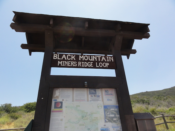
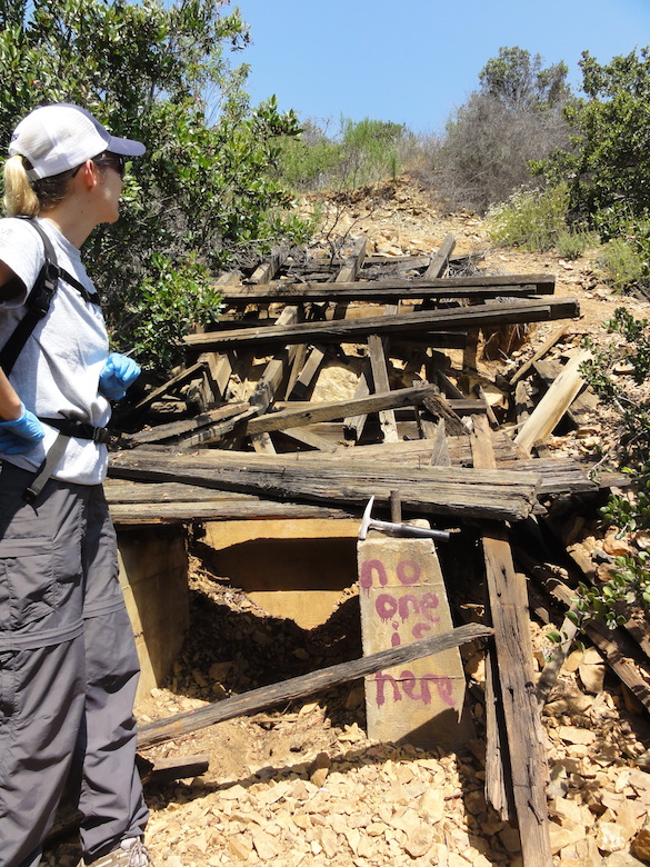
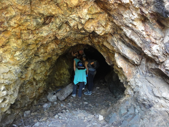
Wooster, Ohio— On April 23, 2016, the Sedimentology and Stratigraphy class took a field trip to the local Wooster Memorial Park, also called Spangler Park. The goal was to study three separate outcrops, and then do a little exploring of our own.
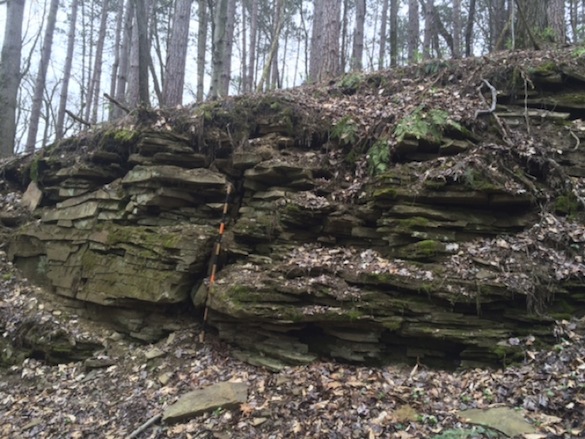
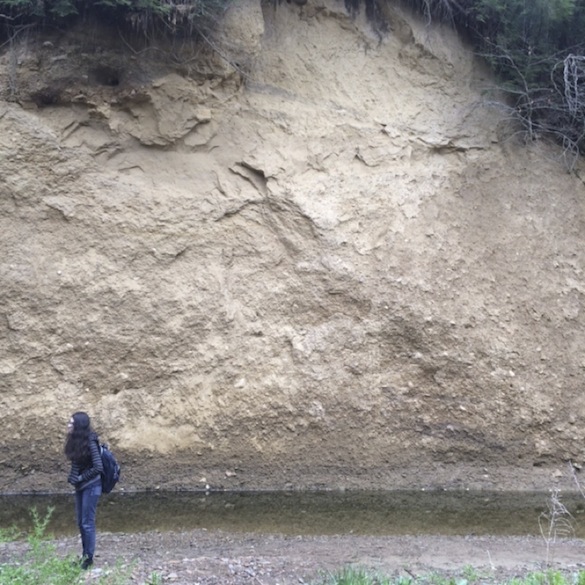
The first stop was a short walk from the entrance to the park, specifically at 40.81475° North and 82.02383° West (above).
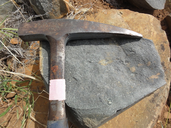
This outcrop contains rocks from the Logan Formation of the Lower Carboniferous. The rocks were non-laminated and of silt size, so it is made of siltstone. There are signs of a little bit of oxidation. There are also ripples present on some of the rocks, which is evidence of a shallow water environment. There were gray shale clasts within the siltstone, which were most likely deposited by storm events. The fact that some of the beds are thicker than others is more evidence of storm events because more sediment would have been deposited during storms and thinner beds would have built up during times of less activity. The bedding angles vary throughout the outcrop, also known as cross-stratification, which is more evidence that ripples and dunes were present as part of a flow regime at the time of deposition.
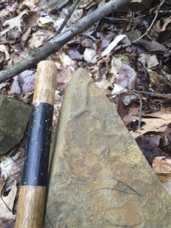 Burrow fossils, which are a form of trace fossil, were left behind by deposit feeding organisms on some of the rocks. This is more evidence of a shallow, marine environment. Based on all the sedimentary structures and characteristics found at this outcrop, these rocks were deposited on the shallow shelf, below the fair weather wave base and above the storm wave base.
The Logan Formation is made up of five members, but specifically the Byer member is likely exposed here. Layers of fine sandstone and siltstones with shale sometimes inter-bedded characterize the Byer member (Hunt, 2009). Although it isn’t present in the two photos above, another member is usually deposited right below the Byer Member. It is called the Berne Member and it is composed of molasse rock, which is a quartz-rich conglomerate formed when the eroded material from continental collisions gathers in a foreland basin. In this case it is eroded material from the continental collisions that built up the Appalachians. The eroded material was then deposited to the west in the foreland basin that covers Pennsylvania and Ohio.
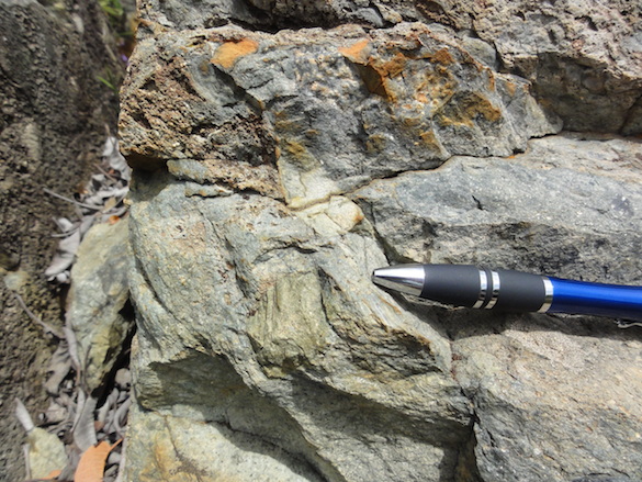
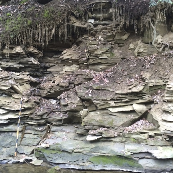
The second outcrop we reached was at the bottom of a gorge, along Rathburn Run, specifically at 40.81784° N and 82.02946° W. The exposure was composed of laminated grey shale from the Cuyahoga Formation. It marked a formation boundary because Logan Formation sandstone lies directly above it. This means the grey shale is older than the Logan Formation. Similar to the Logan Formation, there are trace fossils of marine burrowing organisms within the shale.
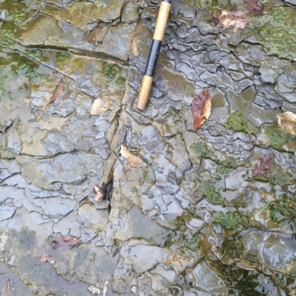 In the above picture you can see an East-West trending joint running through the center of the Cuyahoga Formation grey shale caused by tectonic faulting, which is a phenomenon unrelated to the sedimentary structures.
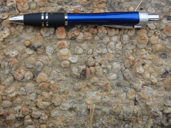
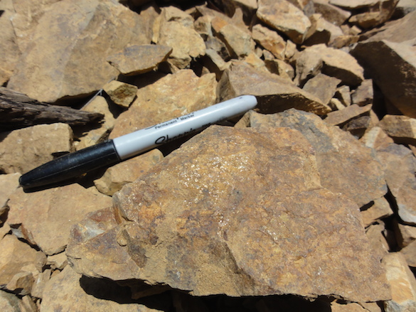
 Siderite deposits were also found in some sandstone at the Rathburn run outcrop, which form after deposition, a diagenetic property. Siderite forms in anoxic environments where iron is reduced and sulfur is present. The grey shale of the Cuyahoga Formation isn’t porous enough for siderite replacement to take place, but the sandstone is.
The third outcrop was father upstream along on a cut bank, located at 40.81903° N and 82.02953° W.
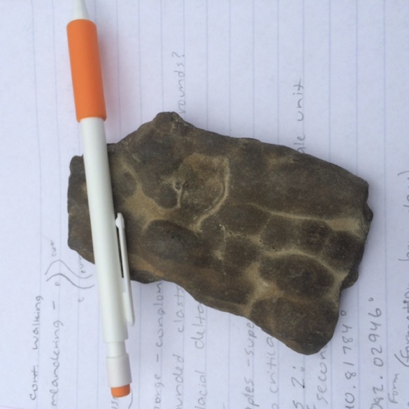
 This photo is taken from across Rathburn Run, from the point bar. This outcrop is much younger in age, from the last time Ohio was affected by glaciation. During the Last Glacial Maximum, specifically the Pleistocene, glacial debris flows deposited the bottom section of the outcrop. The sediment is characterized by a fining upwards sequence and has two scales of support. Some areas of the deposit are composed of large grains within a matrix-support due to debris flow. Other areas of the deposit are composed of sandy conglomerate rock that is grain supported. Overall the sediment is poorly sorted and contains glacial erratics within the sediment, including boulders made of gneiss, granite, and some sedimentary rocks.
A channel cut through the original glacial debris flow deposit and was eventually filled in by wind-blown silt, also known as loess. Loess is characteristically different from the glacial deposit at the bottom of the outcrop. Loess breaks in sheets, which causes it to have steep angles. Overall, the history of this outcrop is that approximately 15,000 years ago debris flow events deposited the glacial sediment at the bottom of the outcrop, then a channel cut into the deposit and that channel eventually filled with eolian (wind-blown) silt.
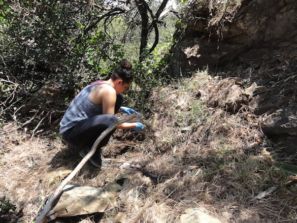
 After venturing a little on our own, a few other students and myself came across a fourth outcrop that was from the Logan Formation at an elevation above both the Cuyahoga Formation shales and the glacial deposits. There is more evidence of jointing and cross-stratification that can be seen in the picture.
We saw two separate formations from the Lower Carboniferous during the field trip. We also were able to see another type of sedimentary deposit that was glacial and eolian in origin. Spangler Park displays and exposes a variety of sedimentary structures and sedimentary characteristics. The park can be characterized as displaying a coarsening upwards sequence with the Cuyahoga shale at the bottom, followed by the coarser siltstone and sandstone of the Logan Formation. This kind of coarsening upwards is usually evidence of either regression or progradation.
Both the Logan and Cuyahoga Formations are representative of shallow marine environments, as was seen in the evidence found at Spangler. Further research shows that the Cuyahoga Formation was deposited as part of a marine environment where the shoreline was prograding during the Kinderhookian and possibly very early Osagean (Bork and Malcuit, 1979; Matchen and Kammer, 2006). The Logan Formation followed and was deposited within a marine proximal deltaic environment during the Osagean (Hunt, 2009; Matchen and Kammer, 2006). This explains the coarsening upwards sequence and the marine sedimentary structures and fossils seen throughout the field trip.
References:
Bork, K.B., and Malcuit, R., 1979, Paleoenvironments of the Cuyahoga and Logan Formations (Mississippian) of central Ohio: Geological Society of America Bulletin II, v. 90, p. 1782-1838.
Hunt, H., 2009, Paleocommunities and Paleoenvironments of the Logan Formation (Mississippian, Osagean) of northeastern Ohio [Undergraduate thesis]: Wooster, The College of Wooster, 50 p.
Matchen, D.L., and Kammer, T.W., 2006, Incised valley fill interpretation for Mississippian Black Hand Sandstone, Appalachian Basin, USA: Implications for glacial eustasy at Kinderhookian-Osagean (Tn2-Tn3) boundary: Sedimentary Geology, v. 191, 89-113.


 Wooster, Ohio — The College of Wooster community will soon say goodbye to Mateer Hall (above), which has housed the Biology Department for decades. It will be demolished next month to make way for the new Ruth Williams Hall of Life Science. I haven’t heard anyone yet say they will miss the creaky and undersized Mateer. The new Life Sciences building, which will be joined to the existing Severance Hall (chemistry), will be beautiful, spacious, and filled with the finest of scientific equipment and facilities.
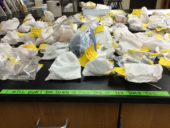
 In the meantime the biologists (sensu lato, including neurobiologists, biochemists and so on) have to go somewhere with all their stuff for two years. Scovel Hall will be home for some of the biology labs, so the geologists have been making room throughout the building. I thought I’d record the process at its most chaotic in Scovel 216 (above) and Scovel 219 (below). The biologists have to move everything out of Mateer in just a few days, so our lab tables and just about every other flat surface in Scovel is occupied by specimens, equipment, and massive bottles of distilled water. I especially like the stuffed animals (including a small bear), the crocodile skulls, and the human skeleton in an ancient tall display cabinet.
 We are looking forward to spending quality time with our biologist friends. We’re each going to learn a great deal about how the other group works, and we’ll have new appreciation for our disciplines. Science marches forward!
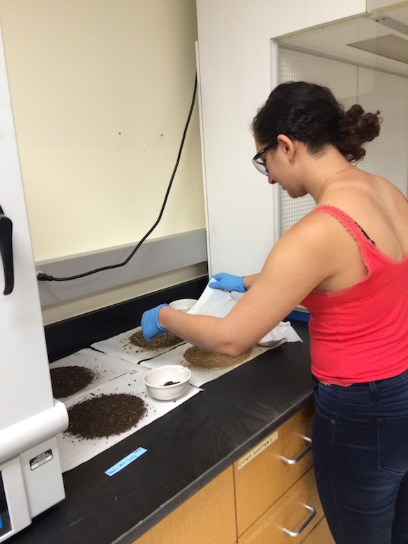
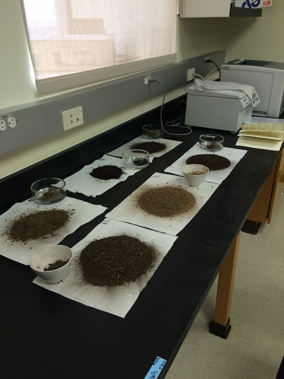
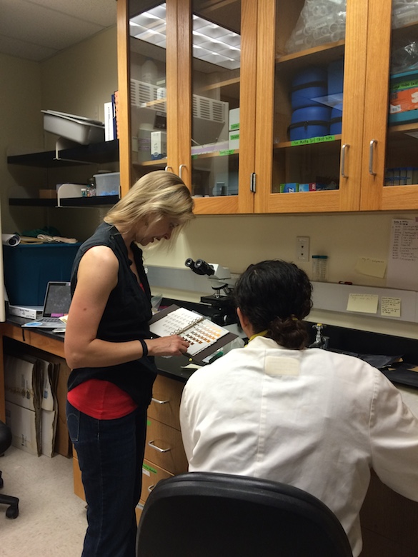
 While her samples dry, Amineh is helping prepare samples for analysis on the scanning electron microscope (SEM-EDS). Doesn’t she look like a happy geochemist?