A Wooster geologist’s summer research experience in The Bahamas: Sarah Bender (’15) and climate and sea level change over the past 6,000 years

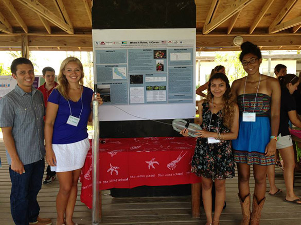
SB coverSarah Bender (’15) and Sarah Frederick (’15) had the opportunity this summer to complete National Science Foundation funded Research Experiences for Undergraduates (REUs). Each spent a good part of their summer completing a research project under the mentorship of accomplished and enthusiastic geologists. Sarah Bender (on the left above) worked under the mentorship of Dr. Lisa Park Boush (’88, center in the photo), a geology professor at the University of Akron, and Kristina Brady (’03, on the right), a curator at the University of Minnesota. A Wooster geology team! This is Sarah’s summer research story in her words and images. (Sarah Frederick’s story is in the previous post.)

This summer I had the pleasure of working with a group of seven interns and four mentors on Eleuthera Island, Bahamas and at the University of Minnesota Twin Cities and Duluth. For two weeks at the beginning of June, we cored three Bahamian lakes, two being blue holes and the other a coastal pond. The goal of this Research Experience for Undergraduates (REU) was to determine the anthropogenic changes that took place in the past thousands of years in the Bahamas by using proxy data from these lakes. The project was led by a Wooster graduate, Dr. Lisa Park Boush (’88), who like myself, was one of Dr. Mark Wilson’s advisees. One of the other mentors, Kristina Brady, also graduated from Wooster (2003) as Dr. Wiles’ advisee, and is now working at LacCore at the University of Minnesota as a curator.

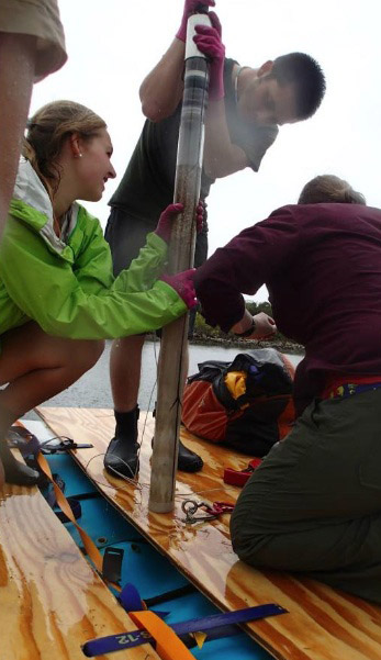
My team worked on the first blue hole, which we named Duck Pond Blue Hole. Duck Pond Blue Hole is an inland circular body of brackish water located in the southern tip of Eleuthera Island. We hypothesize that there are underground conduits connecting the blue hole to the ocean due to the salinity and the fact that the water level was affected by tides. Cores were taken with hand-operated corers from three different spots along a transect of the lake. Overall, my team recovered over four meters of sediment from the three sites! We also took bathymetry data, depth profiles, and did a vegetation survey around the perimeter of Duck Pond Blue Hole.
SB-1a

Myself, a teammate, and Kristina Brady (’03) capping a core from Duck Pond Blue Hole. Check out our mighty coring vessel!

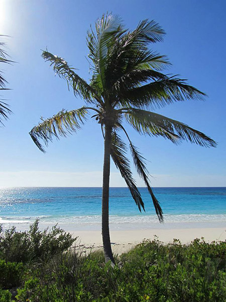
The other team of interns worked on a coastal pond, located directly behind one of the most beautiful beaches in the world. They cored the pond at three sites and took similar lake profiling data as my team. They also worked on dune profiles with Dr. Ilya Buynevich from Temple University using his GPR machine.
SB-2

The other team of interns worked on a coastal pond, located directly behind one of the most beautiful beaches in the world. They cored the pond at three sites and took similar lake profiling data as my team. They also worked on dune profiles with Dr. Ilya Buynevich from Temple University using his GPR machine.

The rest of the time on Eleuthera was spent exploring the island and learning about its history. We took two day-long field trips in which we saw many geological features as well as archaeological sites. With the help of Dr. Perry Gnivecki and Dr. Mary Jane Berman, both from Miami University, we learned all about the native inhabitants of the Bahamas, the Lucayans. We hope our project will help them understand how they were affected by climate change and the landing of Columbus in 1492. Finally, we got to present our preliminary results to the people of the Bahamas at the Cape Eleuthera Institute.

SB-2.5

My teammates and I presenting Duck Pond Blue Hole at CEI.

After finishing fieldwork, we headed to LacCore, the National Lacustrine Core Repository, at the University of Minnesota in Minneapolis to analyze our data. We logged, split, photographed, and described our cores first. We also did a variety of lab work with core samples such as, carbon-14 dating, SEM, loss on ignition, making smear slides, and shell counts. We also got to work at the Large Lakes Observatory in Duluth, Minnesota using the XRF machine and doing grain size analysis.

SB-3

Myself and a teammate prepping samples for grain size analysis at the Large Lakes Observatory in Duluth.

This lab work took about six weeks to complete and we got some amazing results from it. We used the last few days in Minnesota to write our abstracts and make posters for upcoming conferences. Each person took one aspect of our project to focus on. My abstract and poster focuses on the mollusk communities of Duck Pond Blue Hole and how they may be an indicator for climate and sea level change in the Bahamas over the past 6,000 years.

In order to discover what we found, you will have to visit my teammates at GSA in Denver in October or AGU in San Francisco in December. I hope to be able to make it to the AGU conference to help present my team’s work, however, I won’t be presenting my individual abstract until the spring at a regional GSA meeting. If you want to read more on the project, check out the REU Bahamas page on Facebook or the daily blog we kept throughout the project. Now, it’s off to Byron Bay, Australia, for me! I hope everyone had a great summer and I wish you all a successful fall semester!

Posted in Uncategorized | Tagged , , , | Leave a comment

A Wooster geologist’s summer research experience in southeast Wisconsin: Sarah Frederick (’15) and the sourcing of molybdenum in groundwater

SF1This summer, Sarah Bender (’15) and Sarah Frederick (’15) had the opportunity to complete National Science Foundation funded Research Experiences for Undergraduates (REUs). Each spent a good part of their summer completing a research project under the mentorship of accomplished and enthusiastic geologists. Sarah Frederick worked under the mentorship of Dr. Timothy Grundl, a professor within the school of Freshwater Sciences and Department of Geosciences at the University of Wisconsin – Milwaukee. This is her summer research story in her words and images.

My project focused on the elevated levels of molybdenum that have been found in the groundwater of portions of southeast Wisconsin. Molybdenum, for those of you who may not be familiar with this metallic element, is an essential nutrient naturally present at low levels within our environment. High concentrations, however, which are often linked to the improper disposal of such waste products as sewage sludge and fly ash, can create agricultural and health complications. Therefore, the Wisconsin Department of Natural Resources is very interested in this excess and recently completed a two-year study of this problem. The DNR found molybdenum concentrations greater than the recommended quality standards in the water of private wells throughout southeastern Wisconsin, however; this study was largely inconclusive as to a source of contamination. This is where my project began.

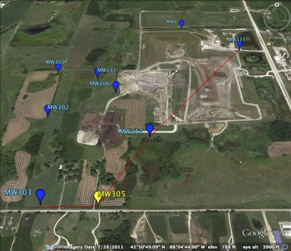
I spent ten weeks investigating the Emerald Park Landfill in Muskego, Wisconsin. The image at the top of this post is an aerial view of Emerald Park. Water samples were collected from the wells marked in blue while the yellow denotes the boring from which soil samples were collected. The red line marks the cross-section I analyzed.

Fieldwork in a landfill was a completely new experience for me. There were two extremes involved. First, near the landfill itself, sampling was a dirty, dusty job and you had to be careful not to get run over by trucks! However, sampling on the outskirts presented its own challenges. As part of this landfill’s wildlife remediation it has created extensive wetland environments. Thus, sampling involved swimming through grass two feet taller than myself in search of wells that one feared may be nonexistent, all while lugging sampling equipment over damp, pitted terrain. In the end we did manage to find all of the wells that we were looking for and successfully collect data and water samples from each.
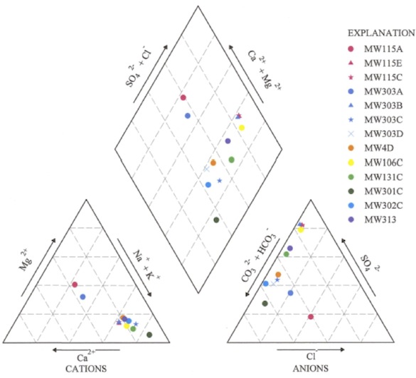
As I was running my samples through the ion chromatography system and the atomic absorption spectrometer back in the laboratory, it quickly became clear that there was a significant difference between the surface water and the water collected from the deeper wells. Piper diagrams clearly illustrated this difference in relative water chemistry. The Piper diagram below displays the relative chemistry of all thirteen of the Emerald Park monitoring wells analyzed. The two A wells, which are shown to be distinct from the rest of the wells, are the wells screened within the surface water.

SF2This hypothesized disconnect between the surface water and the aquifer below was corroborated by the analysis of these water samples using a modeling system called PHREEQC, which was unable to accurately model the transition between the two waters. Further confirmation was provided by the molybdenum concentrations detected within the wells from which a vertical suite of samples was collected. The graph below shows that the surface water contains very little molybdenum while the greatest concentrations appear in the nest deepest wells.

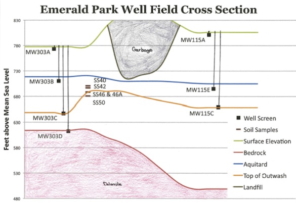
SF3The presence of an aquitard, a dense, impermeable clay hardpan that effectively separates the surface water from the water below, was finally confirmed by the boring logs for this site, which record increased blow counts and a description of this layer. Through the use of well logs and the conclusions of my own research, I thus was able to illustrate the stratigraphy of this site.

SF4The existence of this aquitard and the absence of elevated molybdenum levels within the surface water eliminated the Emerald Park Landfill as a source of contamination since the lowest extent of the landfill storage does not penetrate this clay barrier.

My study therefore concluded that the molybdenum is sourced in the clay. Since almost all the molybdenum detected was dissolved, it was not being transported into the site on colloids. Instead, molybdenum sorbed to hydrous ferric oxides and the cation exchange sites of the clay is being released through reductive dissolution or increased sorption competition.

While this project was only a case study, it is my hope that the Wisconsin DNR will further investigate the possibility that the molybdenum is naturally from the clay of southeast Wisconsin, and that elevated groundwater molybdenum concentrations are a result of the water chemistry.

Posted in Uncategorized | Tagged , , , , | 1 Comment

Wooster’s Fossils of the Week: A foraminiferal ooze from the Pleistocene of Italy

YCM forams 1On a recent field trip to Sicily, our paleontological party visited outcrops at Cala Sant’Antonino on the western side of the Milazzo Peninsula in the northwestern part of the island. We saw there an Early Pleistocene sedimentary unit informally called the “Yellow Calcareous Marls”. With a handlens you would see a close view of the rock like the image above. It consists almost entirely of tiny hollow white spheres with occasional dark flecks. In the lab back home these little calcitic balls were revealed to be tests (skeletons) of foraminiferans known as Globorotalia inflata (d’Orbigny, 1839). This is a classic example of a biogenic sediment called foraminiferal ooze, samples of which are now in Wooster’s paleontological and sedimentological teaching collections.
Foram-Marl-060913This is the outcrop of the “Yellow Calcareous Marls” at Cala Sant’Antonino from which the above samples were collected. The rock is very soft and powdery to the touch.

YCM forams 2In this closer view of the rock the individual foraminiferal tests are more apparent. Near the center is one example showing the connected bulbous chambers (making it multilocular) and the slit-like aperture between them. These tests are slightly recrystallized, giving them a sugary look. The dark bits are sand-sized volcaniclastic grains derived from early eruptions of the Mount Etna complex.

Globorotalia_inflataThese are modern examples of Globorotalia inflata. (The scale bars are 0.1 mm.) The bumpy surface texture, bulbous chambers and distinctive aperture make identification of the fossil examples fairly easy. The images were taken by Bruce Hayward.

Globorotalia inflata is a long-lived planktonic species, meaning it floats about near the top of the water column throughout the oceans. In life these single-celled organisms extend thin strands of material (pseudopodia) into the water around them to collect organic material and the occasional diatom or radiolarian for nutrition. They live in populations with billions of individuals, so under the right conditions their tests can accumulate on the seafloor in numbers so vast they form thick deposits, our foraminiferal oozes. Our particular ooze in this story formed in relatively deep (epibathyal), cool waters during one of the early glacial intervals. This foraminiferan turns out to be a critical guide to the age of the unit as well as its paleoenvironmental context.

References:

Fois, E. 1990. Stratigraphy and palaeogeography of the Capo Milazzo area (NE Sicily, Italy): clues to the evolution of the southern margin of the Tyrrhenian Basin during the Neogene. Palaeogeography, Palaeoclimatology, Palaeoecology 78: 87–108.

Sciuto, F. 2012. Bythocythere solisdeus n. sp. and Cytheropteron eleonorae n. sp. (Crustacea, Ostracoda) from the Early Pleistocene bathyal sediments of Cape Milazzo (NE, Sicily). Geosciences 2012 2: 147-156.


Posted in Uncategorized | Tagged , , , , | Leave a comment

Checking in from the Far East

We are currently finishing our first leg of field research on Sakhalin Island, Fareast Russia, and today we are traveling to Vladivostok to stage the next two weeks of sampling climate-sensitive trees. This is  collaborative Wooster project funded by NSF with Kevin Anchukaitis (Woods Hole Oceanographic Institute) and Rosanne D’Arrigo (Lamont-Doherty Earth Observatory). Our Russian collaborators include Olga Solomina (Russian Academy of Sciences), researchers Ekaterina Dolgova; Eugenio Grabenko Vladimir Matskovsky, Tatiana Maratovna Kouderina and our host on Sakhalin, Yury Gensiorovskiy. Future Wooster student projects will include work on the Kamchatka Peninsula, the Sikhote-Atlin Mountains and the Kurile Islands.

team_dinner

The team at our final dinner at the Far East Branch of Geological Institute in Yuhzno-Sakhalinsk.

victor

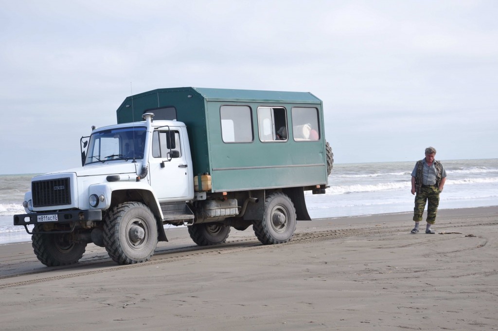
The group split into two teams to find old and climate sensitive trees on the Island. My group traveled with Victor (above) who ably drove us in the Gas66. Here Victor takes a break on the shore of the Sea of Ohotsk.


tatiana_coring

Tatiana (originally from Kazakstan) cores a an old larch in a sea of Pinus Pumulus. This site is on the northern most part of the island – the Smit Peninsula.

camo

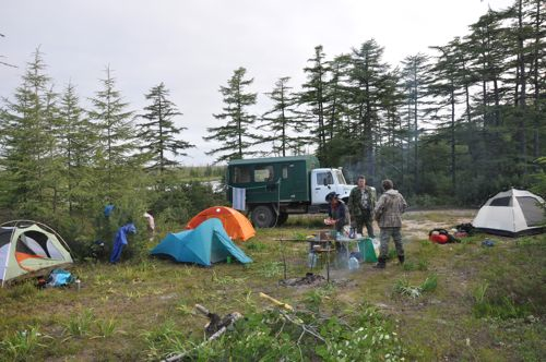
Camp near Nogliki. Olga and I sampled the larch site near here ten years ago and the group updated this important site by re-coring the trees.

strong

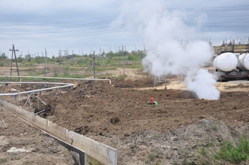
This view is of the many pump jacks and oil wells near Oxa. There are many strong landscapes on the island attesting to an extreme history of logging, oil and gas, fire and political upheaval. In spite of this there are many pockets of old growth forests remaining in beautiful settings.

food

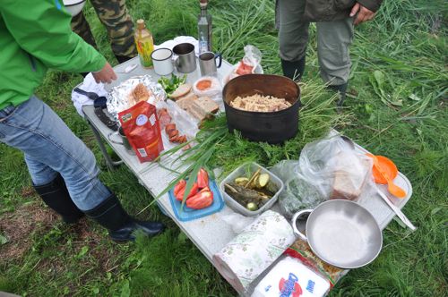
The large of local foods including a full range of sea food makes for excellent dinners after a long day.


Posted in Uncategorized | Tagged , , , | 1 Comment

Wooster Geologist in the Far East of Russia — and on Russian TV!

Screen Shot 2013-08-14 at 9.53.27 AMDr. Greg Wiles, the Ross K. Shoolroy Chair of Natural Resources at Wooster, is currently on an adventurous dendrochronology research trip to the Far East of Russia, including Sakhalin Island. He will have much more to say about it on this blog when he gets the chance. In the meantime, his wife Theresa Ford sent us this link to a Russian news video about his team and their work. The connection is awkward — the video only works for me on my Safari browser — but it is worth the download time to see our Dr. Wiles explaining those wiggly lines and soda straws filled with wood.

There is also a summer 2004 story in Go Nomad touching on Greg’s previous expedition to Sakhalin Island. Theresa found this too, and it was new to me. Here’s a link to a Russian Academy of Sciences page about that earlier research. It has some nice photographs.

Posted in Uncategorized | Tagged , , , | 1 Comment

Wooster’s Fossil of the Week: An almost planispiral gastropod from the Middle Jurassic of southern Israel


Discohelix tunisiensis apical copyAdd this to the list of fossils that have confused me. This summer, during a Wooster expedition, Lizzie Reinthal and Steph Bosch collected the above specimen from the Matmor Formation (Middle Jurassic, Callovian) of southern Israel. I simply assumed it was an ammonite, especially because we were anxious to find ammonites to further reinforce our biostratigraphic framework (how we tell in which geological time interval our fossils belong). When I later tried to identify it by searching through the Jurassic ammonite literature, though, I could find nothing like it. I then sent a photograph to my friend Zeev Lewy, a prominent ammonite expert recently retired from the Geological Survey of Israel. His answer was a surprise: this fossil is the gastropod Discohelix tunisiensis Cox 1969.
Discohelix tunisiensis adapicalHow could this be a snail when it looks so much like a cool, multi-whorled planispiral ammonite, complete with ribs? Well, it is not planispiral, now that I look at it again. Above you see the other side of the specimen, with its slightly depressed center. Most ammonites don’t show such asymmetry. This actually is a gastropod, and it represents an ancient group (the clade Vetigastropoda — don’t get me started on the complications of gastropod systematics!) with primitive features reminiscent of Paleozoic marine snails (from a group I learned to call “archaeogastropods“). It is not as much that the snail has converged on an ammonite style of shell, it’s that the ammonites developed a similar shell much later for entirely different reasons (swimming, for example). Discohelix was likely an herbivore grazing in patchy coral reefs like we have represented in the Matmor Formation. It has become a useful index fossil for the Jurassic of the Tethyan Realm, although this is the first time I’ve found it in Israel.
Pseudotorinia (Architae-group) retiferaThe above is the marine snail Pseudotorinia (Architae-group) retifera. It used to be called Discohelix retifera, and you can see why. It may not be in the same genus, but you can see that this modern group and Discohelix are closely related. Discohelix itself is now known only from the fossil record.
DunkerDiscohelix was named as a genus in 1847 by Wilhelm Bernhard Rudolph Hadrian Dunker (1809-1885), a German natural scientist with interests in geology, paleontology and marine zoology. (I love that middle name of “Hadrian”.) Like so many 19th Century paleontologists, Dunker started with a practical training in mining engineering and then followed a passion for fossils and modern shells. He had a huge collection of materials that eventually ended up in the Museum für Naturkunde in Berlin. He traded and corresponded with many top scientists of his day, including Charles Darwin. He also published many monographs on modern and fossil molluscan taxa. In 1846, he and Hermann von Meyer established the journal Palaeontographica. This journal survives to this day in two descendants: Palaeontographica A (Paleozoology, Stratigraphy) and Palaeontographica B (Paleobotany).

References:

Cox, L.R. 1969. Gasteropodes Jurassiques du Sud-Est Tunisien [Jurassic gastropods from SE Tunisia]. Annales de Paleontologie, Invertebres 55: 241-268.

Grundel, J. 2005. The genus Discohelix Dunker, 1847 (Gastropoda) and on the content of the Discohelicidae Schroder, 1995. Neues Jahrbuch fur Geologie und Palaontologie-Monatshefte 12: 729-748.

Tëmkin, I., Glaubrecht, M. and Köhler, F. 2009. Wilhelm Dunker, his collection, and pteriid systematics. Malacologia 51: 39-79.

Wendt, J.1968. Discohelix (Archaeogastropoda, Euomphalacea) as an index fossil in the Tethyan Jurassic. Palaeontology 11: 554-575.

Posted in Uncategorized | Tagged , , , | 1 Comment

Home Sweet Home…(after 2 months of research and teaching in Utah!!)

WOOSTER, OH — Two months in the field is great for my geologic soul, but I admit that there is an excitement on campus as I prepare for classes to begin in the next few weeks.  I last blogged about my time in Utah weeks ago, when Tricia Hall (’14) and I collected data in central Utah for her I.S. project on deformation bands.  It was difficult for me to blog while teaching field camp in June and July (32 students; 24/7 questions), but I wanted to catch everyone up on some of the sights from this summer.

During our last days in the field together, Tricia and I were both geologists and naturalists, witnessing “survival of the fittest” first-hand.  Check out these action shots:

Snake and Bat 1This snake has caught a bat, which was hiding in one of the fractures in the rock.  Oh…by the way…we just so happened to be taking measurements in this very area!!  As I was taking measurements, my head came within inches of the snake’s rear end.  But, lucky for me, he didn’t see me, as he had shoved his head inside one of the fractures to grab the bat.  Needless to say, when I saw our friend, I broke the world record for the 100 m dash (well, it was more like the steeplechase as I bounded across the rocks).

Snake and Bat 2Our friend ate several bats that afternoon; you can see here that he is busy swallowing one of the bats completely.  But, we still had to grab data, so I sent Tricia back in to get some of the measurements!!  As the diligent advisor, I decided to be “on the look-out” while she took the measurements (placing Tricia between the snake and me).

After my time with deformation bands, I spent time in Ice Springs Volcanic Field with ‘Team Utah 2.0’ (Meagen Pollock, 6 Wooster geology students,  and a group from Albion College led by Thom Wilch).  Meagen did a great job blogging our exploits of our field season, which was definitely enjoyable!!

Then, for the rest of the summer, I taught at Ohio State’s field camp based in Ephraim, Utah, and field camp this year had a record number of students.  While I cannot show you pictures of our mapping areas and tell you about all of the really outstanding geology there (after all, I don’t want to spoil the fun and give away all of the answers for next year’s students), I will say that central Utah has some amazing geology.  The field camp is located in the Sevier fold-thrust belt, and so wonderful foreland basin deposits are the basis of many of our mapping areas.  However, the area has been overprinted by more recent extension, making it a very complex transition between the Basin and Range and the Colorado Plateau.

I would like to share with you some of the really awesome field trips that we took the students on…

Waterpocket MonoclineEarly on, we traveled to Capitol Reef National Park, where the view of the Waterpocket Monocline is phenomenal.  The structure is one of the classic monoclinal folds formed during the Laramide Orogeny.  But, even though I absolutely LOVE monoclines, there was more to see at Capitol Reef…

Capital Reef - Jn Cross-beddingHere is a picture of the Navajo Sandstone and its amazing cross-bedding in all of its glory.  Can you just imagine yourself standing in this large desert environment during the Jurassic?  Picture yourself as a sand grain, saltating along a dune surface…

Capital Reef - GoosenecksBut, I cannot forget to show you a picture from the Goosenecks Overlook in Capitol Reef.  Seeing the stratigraphy in this portion of the part was very helpful to all of the students, as they began to mentally correlate units from southern Utah toward central Utah.

After days of mapping back in central Utah, we took another field trip to Great Basin National Park and the Northern Snake Range (eastern Nevada); this trip with the field camp students is always a highlight for me each summer.

Lehman CavesAt Great Basin National Park, you can take a guided tour of Lehman Caves.  Some of the views inside of the caves are incredible.  The delicate and fragile cave morphologies are spectacular and include stalactites, stalagmites, draperies, shields, and popcorn!!  The added plus to the Lehman Caves tour is that the temperature is always in the 50s, which is such a contrast to the desert heat that I am in all summer.

From Great Basin National Park, we traveled to the Northern Snake Range…

Northern Snake RangeThe Northern Snake Range, seen above, reveals a remarkable metamorphic core complex (MCC).   A MCC is a result of extreme crustal extension, and so you can see highly metamorphosed basement rocks that have been exhumed.

NSRD ScenicAbove is a scenic view of the Northern Snake Range detachment surface (NSRD; note the white rock unit in the picture).  The detachment surface is really a low-angle fault, which reveals metamorphosed rock in the footwall and normal faulted units in the hanging wall.

NSRD FoldingHere is a look at the highly folded metamorphosed rocks of the NSRD.  It literally takes the field camp class hours to walk a transect through all of the rock units leading up to the NSRD, but once they get there, the view is well worth the hike.  This year, we were able to have an amazing view of a forest fire in the Great Basin National Park (Lexington Arch Road wildfire, July 2013).

After a day looking at the NSRD, it was time to examine some other extensional characteristics of this region…

Hendrys CreekTake a look at all of these conjugate, normal faults near the mouth of Hendrys Creek!!  Aren’t they absolutely beautiful?  We were able to take the class up close and personal to these faults, getting accurate measurements for a computer-based exercise for later in the summer.  Students were able to take joint and fault measurements at this locality and foliations and lineations at several other localities within Hendrys Creek.  Then, using Stereonet, they could analyze and interpret the tectonic significance of the area!!  I get to visit Hendrys Creek each summer, and one of my former I.S. students (Joe Wilch ’13) worked in Hendrys as part of his I.S. project with the summer 2012 Keck Geology Consortium.

At the end of the summer, it was back to mapping in central Utah, and this — mapping and teaching mapping — makes me very happy.  I just love to be out in the field.  Each and every day, I get to look at the magnificent Wasatch Monocline with its fantastic Mesozoic-Cenozoic stratigraphy and antithetic normal faults (shown below in a view up Manti Canyon).

Monocline - MCP

IT IS GREAT TO BE A FIELD GEOLOGIST!!


Posted in Uncategorized | Tagged , , , | 4 Comments

Wooster’s Fossil of the Week: An irregular echinoid from the Middle Jurassic of southern Israel

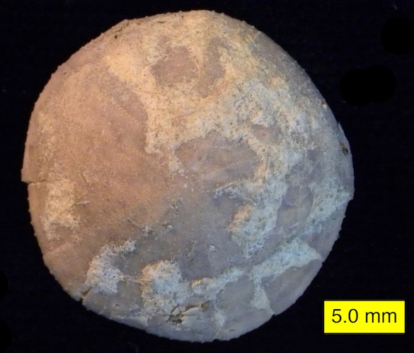
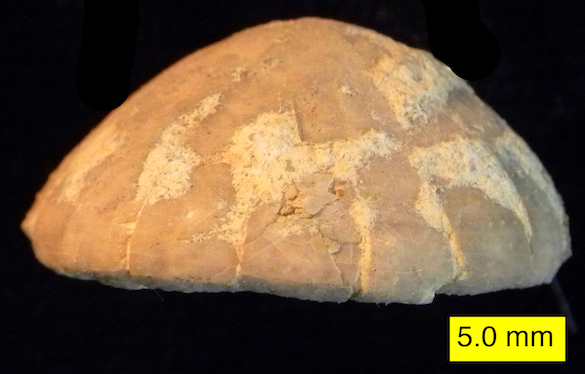
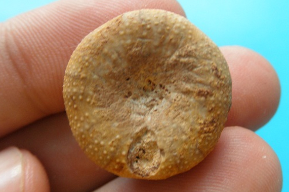
Holectypus depressus adoral 585From the view above, this fossil from the Matmor Formation (Middle Jurassic, Callovian) of southern Israel looks like your standard echinoid (a group that contains sea urchins and sand dollars), but turn it on its side (see below) and you see it is unusual. Echinoids have two large categories: those that are globular in shape (like sea urchins) are called “regular“, and those that are flattened (like sand dollars) are “irregular“. (I know, oddly value-laden terms, these.) This specimen belongs to a group that is rounded on its top portion and flattened on its bottom (oral) surface. This between-ness makes it a fun little specimen.
Holectypus depressus Side 585I could not identify this echinoid, which we collected on our Israel expedition this summer, because I could not find the most important diagnostic features. Fortunately my colleague Andrew Smith, recently retired from the Natural History Museum in London, quickly knew what it was. (This is not surprising — he’s the world’s expert on fossil echinoids. Check out his incredible Echinoid Directory.) Andrew identified this specimen as belonging to the genus Holectypus Desor, 1842, and probably the species Holectypus depressus (Leske, 1778).
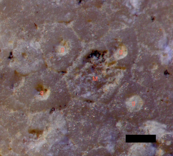
Holectypus depressus apical system 585The first feature Andrew noticed was the apical disk on the very top of the test (the term for an echinoid skeleton). In the above image (where the black scale bar = 200 microns) I’ve labelled the four gonopores (where gametes exit, as you might have guessed) and the madreporite (a sieve plate at the opening of the water vascular system). This arrangement is characteristic of the genus.
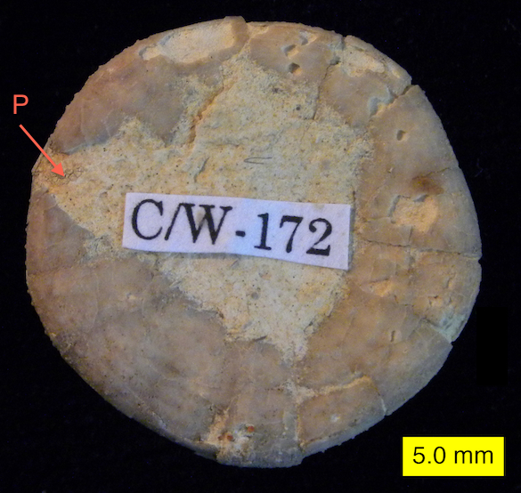
Holectypus depressus oral 585Most surprising to me was Andrew’s identification of the most obvious defining feature of Holectypus, the periproct (the anal opening). I couldn’t find it, but Andrew knew where to look. In this view of the oral surface, it is the gap labeled “P”. Looks just like a place where the test is broken, right?
Callovian France HolectypusHere is the oral surface of an unbroken Holectypus specimen from the Callovian of France. The large periproct is immediately visible as the whole at the bottom. Now the broken margin of the periproct on our specimen makes sense.

Holectypus belongs to a group of irregular echinoids still around today. They are sometimes characterized as having “conservative” evolution, meaning they have not changed much over long periods. The irregular echinoids appeared earlier in the Jurassic as a modification of their regular ancestors. They became flattened and bilateral, the periproct moved out of the apical disk, their ambulacra (rows of tube feet visible as tiny holes radiating from the apical disk on the top image) pulled up away from the mouth, and their spines were reduced in size and increased in number. These were primarily adaptations for burrowing into the sediment. Holectypus has retained its inflated upper portion, has relatively large spines (some still cling to our specimen), and still is circular in outline. It was a deposit feeder but not specialized for burrowing.

We wouldn’t want to call this a “transitional fossil”, but it is a nice example of the gradient of adaptations present when there is a major outbreak of innovation as during the rise of the irregular echinoids in the Jurassic.

References:

Kroh, A. and Smith, A.B. 2010. The phylogeny and classification of post-Palaeozoic echinoids. Journal of Systematic Palaeontology 8: 147-212.

Rose, E.P.F. and Olver, J.B.S. 1985. Slow evolution in the Holectypidae, a family of primitive irregular echinoids, p. 81-89. In: Keegan, B.F. and O’Connor, B.D.S. (eds.), Proceedings of the Fifth International Echinoderm Conference, Galway, 24-29 September, 1984.

Saucède, T., Mooi, R. and David, B. 2007. Phylogeny and origin of Jurassic irregular echinoids (Echinodermata: Echinoidea). Geological Magazine 144: 333-359.

Posted in Uncategorized | Tagged , , , | Leave a comment

Wooster’s Fossil of the Week: An infected crinoid from the Middle Jurassic of southern Israel

CrinoidGalls03 copyThis weathered beauty is a stem fragment of the articulate crinoid Apiocrinites negevensis from the Matmor Formation (Middle Jurassic, Callovian) of the Negev, southern Israel. The regular divisions you see making up the stem are the columnals, which look a bit like a stack of poker chips. You can even make out the crenulations on the articulating faces of the columnals, seen as tiny zig-zags. What is unusual about this stem, of course, are the large swellings with multiple holes. These appear to be something like the galls you sometimes see in plant stems formed when a parasite is surrounded by living plant tissue.
CrinoidGalls02 copySenior Independent Studies student Lizzie Reinthal (’14) is working on these odd structures (we have dozens of examples) as part of her investigation of the taphonomy of A. negevensis in the Matmor Formation. We know that the swellings were made by the interaction of some sort of organism with the living crinoid, but we don’t yet know the timing or mechanism. It could be that the holes were drilled first into the stem and the crinoid grew the extra skeletal tissue to essentially push them away, or the swellings could have been the equivalent of galls and some sort of enclosed animal bored its way out of the structure. (And an extra point to those of you who spotted the barnacle boring! Note that it has no swelling around it and thus was likely drilled after the death of the crinoid.)

These infected crinoid stems were first described from the Matmor by Feldman and Brett (1998). They suggested they were from parasitic myzostome worms, which are usually found on crinoid arms and have a long fossil record (see Meyer and Ausich, 1983, and Hess, 2010). They could also be from some sort of embedded organism like that represented by Phosphannulus on Paleozoic crinoid stems (Welch, 1976).

Lizzie will be pursuing the mystery by careful sectioning some of these swellings and seeing if she can relate the crinoid skeletal growth patterns to either a borer or an embedded parasite. Unfortunately that means we must destroy some specimens to better understand the phenomenon, a classic dilemma paleontologists sometimes face.

References:

Feldman, H.R. and Brett, C.E. 1998. Epi- and endobiontic organisms on Late Jurassic crinoid columns from the Negev Desert, Israel: Implications for co-evolution. Lethaia 31: 57–71.

Hess, H. 2010. Myzostome deformation on arms of the Early Jurassic crinoid Balanocrinus gracilis (Charlesworth). Journal of Paleontology 84: 1031-1034.

Meyer, D.L. and Ausich, W.I. 1983. Biotic interactions among recent and fossil crinoids, p. 377–427. In: Tevesz, M.J.S. and McCall, P.L., eds., Biotic interactions in recent and fossil benthic communities. Plenum Press, New York.

Welch, J.R. 1976. Phosphannulus on Paleozoic crinoid stems. Journal of Paleontology 50: 218-225.

Posted in Uncategorized | Tagged , , , , | 3 Comments

Science Highlights from IAVCEI 2013

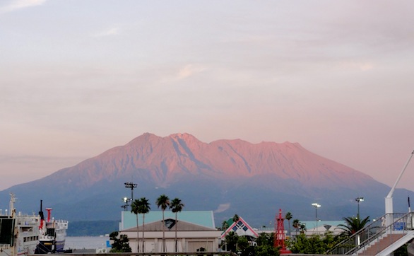
KAGOSHIMA, JAPAN – Although the eruption of Sakurajima on the mid-conference field trip was the ultimate highlight of the IAVCEI 2013 Scientific Assembly, let’s not forget that there were also four full days of excellent talks and posters on the cutting edge of research in volcanology.

Sakurajima at sunset, from Kagoshima.

Sakurajima at sunset, from Kagoshima.

There were far too many inspiring presentations to name them all, so here are just a few scientific highlights:

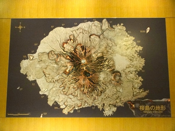
A RRIM of Sakurajima hangs in the Sakurajima Visitor Center.

A RRIM of Sakurajima hangs in the Sakurajima Visitor Center.

Thanks to all ~1,100 international scientists for making my first IAVCEI conference a memorable experience! I look forward to seeing all of you again in 2017, when the next scientific assembly of IAVCEI will be held in Portland, Oregon.

Posted in Uncategorized | Tagged , , , | Leave a comment