


 Readers of this blog will remember Kate Runciman, a 2022 graduate of The College of Wooster and now a graduate student at the University of Cambridge. Her Independent Study thesis (after peer review and revisions) has now been published in a conference volume from the 2022 meeting of the International Bryozoology Association (Runciman et al., 2023). She studied growth patterns within large trepostome bryozoan skeletons from the Cincinnati, Ohio, region. They are from various units in the exposed Upper Ordovician (Katian) rocks.
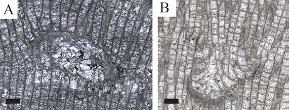
The images above are from acetate peels, with the scale bars equal to 0.50 mm. Figure A shows zooids that grew laterally over a cavity opening, sealing it off with a flat “roof” (C/W-152-2; Amplexopora (?) filiasa). Figure B shows zooids that budded down into a cavity and then angled upwards (C/W-152-1; Amplexopora robusta).
In this new paper, Kate and her colleagues explore the paleoecology of trepostome bryozoans and the various organisms that interacted with them on the Ordovician seafloor. They cover topics from borings, bioclaustrations and parasites to self-overgrowths, brown bodies and “ghosts” of boring inhabitants.
The second paper in the same volume is from a project led by our friend and colleague Caroline Buttler, who is based in the Department of Natural Sciences, Museum Wales, Cardiff. She led a team using non-destructive 3D imaging technology to explore skeletal details of Paleozoic palaeostome bryozoans. The results are spectacular (Buttler et al., 2023). The images obtained by X-ray Micro Computed Tomography (X-ray μCT) and Microscopy (XRM) are have exquisite details.
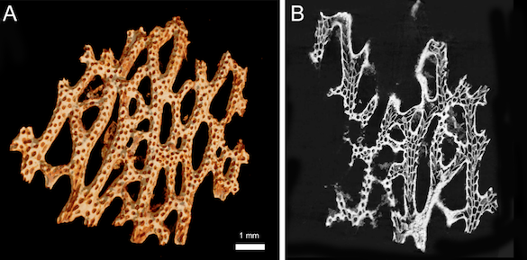 The images above show the silicified fenestrate bryozoan Oeciophylloporina in 3-D (A) and a “digital thin section” (B). Amazing detail. The specimen is from the Lower Silurian (Aeronian) of the Derenjal Mountains in central Iran.
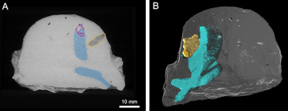 Another example of the application of this X-ray μCT (the part I was directly involved in) was to look at the internal features of borings in hemispherical calcitic trepostome bryozoans. The above images show a bryozoan from the Upper Ordovician (Sandbian) of Estonia (C/W-231-1). In A the borings are shown in transverse section; in B the segmentation of the borings with the rest of colony are made digitally transparent. We’ve just started to explore what these tomography techniques can tell us about the development of these borings, their possible inhabitants, and how the bryozoans responded to them.
Fossil bryozoology advances!
References Cited
Buttler, C.J., Mitchell, R.L., Wilson, M.A. and Johnston, R.E. 2023. Applications for x-ray tomography/microscopy of Palaeozoic palaeostome bryozoans, p. 1–8. In: Key, M.M., Jr., Porter, J.S. and Wyse Jackson, P.N. (eds) Bryozoan Studies 2022. CRC Press/Balkema, Abingdon and Boca Raton.
Runciman, K.M., Wilson, M.A., Buttler, C.J. and Judge, S.A. 2023. Colony repair strategies in large trepostome bryozoans from the Upper Ordovician (Katian) of the Cincinnati region, USA, p. 105–111. In: Key, M.M., Jr., Porter, J.S. and Wyse Jackson, P.N. (eds) Bryozoan Studies 2022. CRC Press/Balkema, Abingdon and Boca Raton.