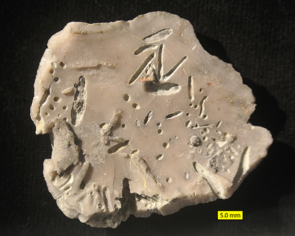
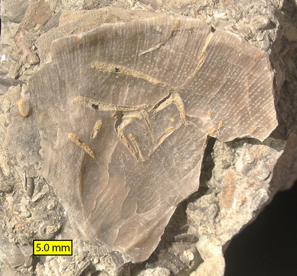
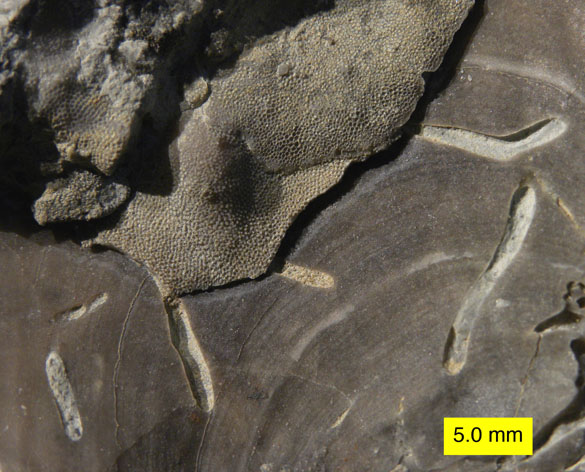
Wooster’s Fossil of the Week: A whale ear bone (Neogene)

Whale inner earbonesThis is another fossil that has sat in a display case for decades in Scovel before I really examined it. Unlike last week’s specimen, though, it has no identifying label on its reverse. This is always a serious disappointment for science — no location! I show the fossil above with a front and back view (as much as there is a front or back). We are looking at an auditory bulla (part of the middle ear system) of an ancient whale. The most we can say is that this may be from a type of sperm whale that lived during the Neogene. Likely this specimen was collected on the east coast of the United States, maybe Maryland or Virginia.

Surprisingly, whale ear bones are rather common in the later fossil record. They seem to have been of denser bone than the rest of the whale skeleton, so they were better preserved. The auditory bulla is a bony cover for the delicate middle ear bones and tissues. In humans it is part of our temporal bone. Whales have several adaptations in their ears for hearing underwater. They have no external ear opening. They use instead the lower jawbone to transmit vibrations to the ear complex (something like what many snakes do). They have a pad of fat to enhance these vibrations for the tiny ear bones (tiny relative to the massive size of the whale). You can learn much more about fossil whale ear bones at this excellent blog post from the Virginia Museum of Natural History.

You are asking, though, fine enough, but how can I use a fossil whale ear bone? There’s a video to train you! These bones have “ancient, ancient memory” that is “preserved sonically”. Just be sure to hold it in your non-dominant hand and remember that “this is an art”. Do it correctly and you will have tapped into the wisdom of our ancient whale brothers and sisters. To think that every day I walked blithely by this portal to the Knowledge of the Ages.

References:

Fraser, F.C. and Purves, P.E. 1960. Hearing in cetaceans: evolution of the accessory air sacs and the structure and function of the outer and middle ear in recent cetaceans. Bulletin of the British Museum (Natural History) 7: 1-140.

Ketten, D.R. 1997. Structure and function in whale ears. Bioacoustics 8: 103-135.

Posted in Uncategorized | Tagged , , | 4 Comments

Wooster paleontologists begin a new field season

Southgate 030814RICHMOND, INDIANA–This is the first day of what upper midwesterners hilariously call “spring break”, so it is time to get some students in the field. I can’t say this is the first Wooster geology fieldwork of the year because that crazy Greg Wiles lab was out on the ice in deepest January. I spent today in eastern Indiana exploring field sites for a new generation of Independent Study students. Tomorrow and Monday Coleman Fitch (’15) and William Harrison (’15) will be joining me to collect specimens for their I.S. projects on Cincinnatian (Upper Ordovician) fossils. We’ll highlight their work in the next couple of days.

Above is one of the best known fossil sites in southeastern Indiana. It is the Southgate Hill section (sometimes called the St. Leon roadcut) at N 39.33899°, W 84.95287°. Exposed here are (from bottom to top) the Oregonia, Waynesville, Liberty, Whitewater and Saluda units of the Cincinnatian Group. It is a rich site — and incredibly muddy today. I suppose I’ll take mud over ice. Note the blue sky. By the end of the day it was as gray as the rocks, making the search for tiny fossil details difficult. Tomorrow promises to be much sunnier. Brach Slab 030814The brachiopods at the Southgate exposure are incredibly abundant and well preserved. These are strophomenids. Crinoids Bryozoans 030814Bryozoans (the twiggy bits) and crinoids (the circular fossils with star-shaped central holes). Can’t go wrong with this combination. More tomorrow and Monday as Coleman and William get to work. Meanwhile I’m wondering how I managed to get a motel room right next to an active railway …

Posted in Uncategorized | Tagged , , | 2 Comments

Wooster’s Fossil of the Week: An agate-replaced coral from the Oligocene-Miocene of Florida

DSC_3384_585I long thought of this beautiful specimen as more rock than fossil. It is a scleractinian coral that has had its outer skeleton replaced by the silicate material agate and its interior skeleton completely hollowed out. The result is a geode that happens to also be a fossil.
FLMNH_585Then during last month’s North American Paleontological Convention in Gainesville, Florida, I saw the above specimens on display in the Florida Museum of Natural History. These fossils were so striking that I decided to highlight our single example.
DSC_3388_585This is a view of the top surface of the Wooster specimen. In the upper left is an array of holes with crystals radiating away from them. These are remnants of the original corallites, and there is just enough information there for us to conclude the likely genus is Montastraea. This piece thus becomes an example of Florida’s official state stone. Here’s the official definition: “… a chalcedony pseudomorph after coral, appearing as limestone geodes lined with botryoidal agate or quartz crystals and drusy quartz fingers, indigenous to Florida.” Our specimen came from the Hawthorn Group of rocks near Tampa, Florida.
DSC_3393_585The outside of the fossil shows horizontal banding remaining from the original growth lines in the coral, which is another clue that this is Montastraea. The coral made its skeleton of aragonite around 30 million years ago. After death and burial, silica-rich groundwater began to replace the aragonite on the surface of the coral with what later became banded agate. The interior dissolved away into a hollow cavity.

The common name for this fossil is “agatized coral“, and it is a collector’s item. It is apparently Florida’s only native gemstone. Pretty cool that their state rock and gemstone is a fossil!

References:

Scott, T.M. 1990. The lithostratigraphy of the Hawthorn Group of peninsular Florida. World Phosphate Deposits 3: 325-336.

Posted in Uncategorized | Tagged , , , , | 2 Comments

Wooster’s Fossil of the Week: An interlocking rugose and tabulate coral (Devonian of Michigan)

Hexagonaria percarinata colony viewThis beautifully polished fossil looks like half of an antique bowling ball. Normally I hate polished fossils because the external details have been erased, but in this case the smooth surface reveals details about the organisms and their relationship. We have here a large colonial rugose coral with a smaller tabulate coral embedded within it. The specimen is from the Devonian of Michigan. It may look familiar because it is a large “Petoskey Stone“, the state stone (not fossil!) of Michigan. The large rugose coral is Hexagonaria percarinata (Sloss, 1939).
Hexagonaria percarinata close view 585In this closer view you can see the multiple star-like corallites of this coral. Each corallite held a tentacular feeding polyp in life. The radiating lines are thin vertical sheets of skeleton called septa. The corallites in this type of coral shared common walls and nestled up against each other as close as possible. In the lower center of the image you can see a very small corallite that represents a newly-budded polyp inserting itself as the colony grew. If rugose corals were like modern corals (and they probably were), the polyps were little sessile benthic carnivores catching small passing organisms with a set of tentacles. They may also have had photosymbionts to provide oxygen and carbohydrates through photosynthesis.
Tabulate coral intergrown with HexagonariaIn the midst of the rugose coral is this irregular patch with another type of coral: a tabulate coral distinguished by numerous horizontal partitions in its corallites (and no septa). It was likely a favositid coral, sometimes called a “honeycomb coral”. It was clearly living in the rugosan skeleton and not pushed into it by later burial. Note, though, the ragged boundary between the two corals. The rugose coral has the worst of it with some corallites deeply eroded. What seems to have happened is that the rugose coral had an irregular opening in its corallum (colonial skeleton) after death and the tabulate grew within the space, eventually filling it. The tabulate likely stuck out far above the rugose perimeter, but the polishing shaved them down to the same level. This is thus not a symbiotic relationship but one that happened after the death of the rugose coral. [See comments, however!]
Stumm, Erwin C copyThe rugose coral species, Hexagonaria percarinata, was named in 1939 by Laurence Sloss, a famous sedimentary geologist with an early start in paleontology, but it is best known through the research of Erwin Charles Stumm (1908-1969; pictured above). Stumm was at the end of his life a Professor of Geology and Mineralogy and the Curator of Paleozoic Invertebrates in the Museum of Paleontology at the University of Michigan. Stumm grew up in California and then moved east for his college (George Washington University, ’32) and graduate (PhD from Princeton in 1936) education. He taught geology at Oberlin College up the road for ten years, and then moved to Michigan to start as an Associate Curator and Assistant Professor. I knew his name because in 1967 he was President of the Paleontological Society. He is said to have been a dedicated teacher of undergraduates and effective graduate advisor. It is fitting that his name is connected to such a popular fossil as Hexagonaria percarinata.

References:

Sloss, L. 1939. Devonian rugose corals from the Traverse Beds of Michigan. Journal of Paleontology 13: 52-73.

Stumm, E.C. 1967. Growth stages in the Middle Devonian rugose coral species Hexagonaria anna (Whitfield) from the Traverse Group of Michigan. Contributions from the Museum of Paleontology, The University of Michigan 21(5): 105-108.

Stumm, E.C. 1970. Corals of the Traverse Group of Michigan Part 13, Hexagonaria. Contributions from the Museum of Paleontology, The University of Michigan 23(5): 81-91.

Posted in Uncategorized | Tagged , , , | 4 Comments

A visit to the Florida Museum of Natural History

1 Steph Lizzie Ground Sloth 021714GAINESVILLE, FLORIDA — The 10th North American Paleontological Convention here is sponsored by the Florida Museum of Natural History. (The meeting is excellent, by the way, and very well organized. Congratulations to the paleontological team that put it together.) Since the public display halls of the museum are just across the street from the convention center, Steph Bosch (’14), Lizzie Reinthal (’14) and I gave it a visit. You can’t go wrong with a natural history museum, but this one was extra fun and informative.

The first section of the museum we explored was the Hall of Florida Fossils. For a state with what I thought was a limited set of rocky outcrops and geological units, the range of Florida fossils is extraordinary. They are displayed here from microfossils (shown as magnified models) to large mammal skeletons. Steph and Lizzie are shown above looking up at a gynormous ground sloth skeleton from the Pleistocene.

2 Lizzie Steph Shark 021714A star of the Florida fossil exhibit is a reconstructed set of jaws from the giant Neogene shark Carcharodon megalodon. It has hundreds of  original teeth in several rows. I’m sure this photo setting has been used thousands of times by now.

3 Rainforest Visual 021714My favorite part of the museum was the Butterfly Rainforest. It is a large screened enclosure with thousands of live butterflies, and several birds as well, with dozens of tropical plant species. I liked it so much I went through it twice. (The students were somewhat less enthusiastic.) Butterflies flitted about, often landing in sunny spots and displaying their brilliant colors and patterns. (Yes, this means colorful photographs follow!)

4 Morpho chrysalis 021714 585Just before you enter the enclosure there is a nursery for rearing butterflies, from caterpillar to chrysalis to newly-emerged adult. Above is a specimen of Morpho peleides (Common or Blue Morpho) still clinging to the husk of its chrysalis. Several siblings wait for their unveiling.

5 Morpho on guidebookAfter we entered the enclosure, I opened an outdoor guidebook to identify the butterflies. Maybe it was just chance, or maybe the attraction of the photograph, but a rather worn Morpho peleides landed right on its  species description.

6 Geometric butterfly 021714 585I could not identify the other butterflies I photographed, but that won’t stop me from showing a couple more images. I like the geometric patterns on the wings of this species.

7 Two butterflies 021714This divergent pair shared a patch of sunlight. If any reader happens to know the names of these butterflies, please add them to the comments and I’ll update this text. [Katherine Marenco kindly identified the orange butterfly above as a Gulf Fritillary (Agraulis vanillae).]

There is much more at the Florida Museum of Natural History than we could see in our brief time there. We highly recommend if you’re in Gainesville that you visit this wonderful, friendly, enriching complex.


Posted in Uncategorized | Tagged , , | 2 Comments

Wooster Geologists at the North American Paleontological Convention in Florida

Lizzie & Steph 021514GAINESVILLE, FLORIDA–Steph Bosch (’14), Lizzie Reinthal (’14) and I flew out of icy Ohio this weekend to attend the 10th North American Paleontological Convention in warm, sunny northern Florida. The students jointly presented the beautiful poster above on their Independent Study projects in the Matmor Formation (Middle Jurassic, Callovian) of southern Israel. It was very well received, especially with the addition of fantastic scanning electron microscope images of bryozoans produced by our colleague Paul Taylor at the Natural History Museum in London.

Crowd scene 021514Here’s a crowd scene from the first poster session at NAPC. If you look closely in the center, you’ll see two Wooster alumnae who are prominent paleontologists. Can’t swing a cat at a paleo meeting without hitting Wooster Geologists.

Hilton 021514This is a nondescript image of our hotel and convention center in Gainesville. I show it only to marvel in the blue, blue sky and perfect temperatures. We are on the University of Florida campus near the Florida Museum of Natural History. The paleontology staff at that museum is sponsoring this meeting — and they are doing an extraordinary job made more complex by the absence of about a third of the participants still snow-bound in the north. We escaped through a window of clear weather in Ohio.

Posted in Uncategorized | Tagged , , , , , | Leave a comment

Wooster’s Fossil of the Week: A tubeworm-encrusted parasitic gastropod (Silurian of Indiana)

Platyostoma1_585Last week three Wooster geology students and I visited Ken Karns, an enthusiastic citizen scientist who has developed an extraordinary fossil collection in his home in Lancaster, Ohio. Ken is a man of prodigious energies and skills as he not only is an expert fossil collector and preparator, he also has a world-class curated collection of Ohio beetles! He was introduced to us by our friend Brian Bade, a man with similar enthusiasms and skills. The students were Steph Bosch (’14), Lizzie Reinthal (’14) and Ian Tulungen (’15). Our goals were to meet Ken, see his magnificent collection with Brian and other friends, and then focus on a project for Ian’s future Independent Study work. Success on all counts, and the specimen above is evidence. Ken was very generous in loaning this specimen to us along with several others for Ian’s work.

The above specimen is from the type section of the Waldron Shale Member (Silurian, Wenlockian, Homerian, about 430 million years old) of the Pleasant Mills Formation near St. Paul, south-central Indiana. Ken Karns collected and prepared it. It is a platyceratid snail of the genus Platyostoma Conrad 1842. It is probably of the species P. niagarense Hall 1852, but there is another species in the same unit (P. plebeium Hall 1876). I’m not quite sure of the differences between these species because platyceratids are notoriously variable. It is possible they are synonymous. Unlike most gastropods, platyceratids had calcite shells instead of aragonite, so they are very well preserved. For an excellent taxonomic review of the genus Platyostoma and its founder, Timothy Abbott Conrad, please see Tony Edger’s blog entry. (We’ve talked about Conrad in this blog as well.)
Platyostoma2_585In this different angle on the specimen you can see additional encrusters (sclerobionts) on the surface of the Platyostoma shell. In the lower right is a remnant of a sheet-like bryozoan, but the most prominent sclerobionts are the tubeworms Cornulites proprius Hall 1876. These encrusters interest us very much.
Cornulitids on Platyostoma_585In this closer view it is apparent that several of the cornulitids are aligned with their apertures pointing in the same way. This is a pattern we’ve seen on many of these snails. Platyostoma was a parasitic snail that lived attached to crinoids, which were abundant in the Waldron fauna. They lived high on the calyx of the crinoid firmly fixed to its skeleton. These cornulitids and other encrusters were thus living high off the substrate perched on the snails. They were filter-feeders like the crinoids, so they may have been feeding on some suspended food fraction missed by the crinoid arms, or they were competing for nutrients and added to the parasitic load on the poor crinoids. The cornulitids were further living on a living snail shell, from what we can tell, so they grew with a substrate slowly growing underneath them. This produces all sorts of delicious paleoecological questions to sort out!
Platyostoma long cornulitid_585Check out the size of this specimen of Cornulites proprius attached to another Platyostoma niagarense. Clearly these tubeworms could do very well under these conditions! This is the largest cornulitid I’ve seen.

Ken_Karns_preparatory_labHere is Ken Karns in his fossil preparation laboratory, which he assembled himself. The box with the armholes is for air-abrading specimens to remove matrix.

Display cases KenThis is one section of the display cases Ken has in his basement museum. Most of the specimens shown here are from the Waldron Shale.

Platyostoma collection displayedA closer view of a display of Platyostoma from the Waldron Shale. Note the many encrusters.

Lizzie Brian KenLizzie Reinthal, Brian Bade and Ken talk about fossil preparation with some Waldron material. The cases are full of curated specimens.

Encrusted crinoid rootsThere are so many treasures in Ken’s collections. I am fascinated by this little slab showing the holdfast of a crinoid with sheet-like bryozoans encrusting it. The bryozoans show that the roots were at least partially exposed at some point.

Thank you again to Brian Bade for arranging this trip, and Ken Karns for being such a fantastic host. We are looking forward to many Waldron projects in the future!

References:

Baumiller, T.K. 2003. Evaluating the interaction between platyceratid gastropods and crinoids: a cost–benefit approach. Palaeogeography, Palaeoclimatology, Palaeoecology 201: 199-209.

Baumiller, T.K. and Gahn, F.J. 2002. Fossil record of parasitism on marine invertebrates with special emphasis on the platyceratid-crinoid interaction. Paleontological Society Papers 8: 195-210.

Brett, C.E., Cramer, B.D., McLaughlin, P.I., Kleffner, M.A., Showers, W.J. and Thomka, J.R. 2012. Revised Telychian–Sheinwoodian (Silurian) stratigraphy of the Laurentian mid-continent: building uniform nomenclature along the Cincinnati Arch. Bulletin of Geosciences 87: 733–753.

Feldman, H.R. 1989. Taphonomic processes in the Waldron Shale, Silurian, southern Indiana. Palaios 4: 144-156.

Gahn, F.J. and Baumiller, T.K. 2006. Using platyceratid gastropod behaviour to test functional morphology. Historical Biology 18: 397-404.

Gahn, F.J., Fabian, A. and Baumiller, T.K. 2003. Additional evidence for the drilling behavior of Paleozoic gastropods. Acta Palaeontologica Polonica 48: 156-156.

Hall, J. 1881. Descriptions of the Species of Fossils Found in the Niagara Group at Waldron, Indiana. In: Indiana Department of Geology and Natural Resources, Eleventh Annual Report, p. 217-345. [PDF of the text downloadable here.]

Liddell, W.D. and Brett, C.E. (1982). Skeletal overgrowths among epizoans from the Silurian (Wenlockian) Waldron Shale. Paleobiology 8: 67-78.

Peters, S.E. and Bork, K.B. 1998. Secondary tiering on crinoids from the Waldron Shale (Silurian: Wenlockian) of Indiana. Journal of Paleontology 72: 887-894.

Sutton, M.D., Briggs, D.E.G., Siveter, D.J. and Siveter, D.J. 2006. Fossilized soft tissues in a Silurian platyceratid gastropod. Proceedings of the Royal Society B: Biological Science 273(1590): 1039-1044.

Taylor, P.D. and Wilson, M.A. 2003. Palaeoecology and evolution of marine hard substrate communities. Earth-Science Reviews 62: 1-103.

Posted in Uncategorized | Tagged , , , | 3 Comments

Wooster’s Fossils of the Week: Bioclaustration-boring structures in bryozoans from the Upper Ordovician of the Cincinnati region

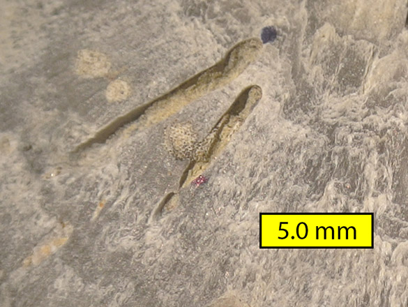
Chimneys 149aAnother bioerosion mystery from those fascinating Upper Ordovician rocks around Cincinnati. Above you see a flat, bifoliate trepostome bryozoan (probably Peronopora) with pock holes scattered across its surface. At first you may think, after reading so many blog posts here, that these are again the simple cylindrical boring Trypanites, but then you note that they are shallow and have raised rims so that they look like little meteorite craters. These holes thus represent tiny organisms on the bryozoan surface while it was alive. The bryozoan grew around these infesters, producing the reaction tissue of the rims. This is a kind of preservation called bioclaustration (literally, “walled-in life” from the same root in claustrophobia and cloisters). The specimen is from locality C/W-149 (Liberty Formation near Brookville, Franklin County, Indiana; 39º 28.847′ N, 84º 56.941′ W).
Chimneys 153aThis is another trepostome bryozoan with these rimmed pits. It is from locality C/W-153 (Bull Fork Formation near Maysville, Mason County, Kentucky; 38º 35.111′ N, 083º 42.094′ W). The pits are more numerous and have more pronounced reaction rims.
Chimneys 153bA closer view. One of the interesting questions is whether these pits are also borings. Did they cut down into the bryozoan skeleton at the same time it was growing up around them? We should be able to answer that by making a cross-section through the pits to see what their bases look like. The bryozoan walls should be either cut or entire.
Chimneys 153cThis is an older image I made back in the days of film to show the density of the rimmed pits in the same bryozoan as above. If we assume that the pit-maker was a filter-feeding organism, how did it affect the nutrient intake of the host bryozoan? Maybe the infester had a larger feeding apparatus and took a larger size fraction of the suspended food? (This could be a project where we apply aerosol filtration theory.)  Maybe the bryozoan suffered from a cut in its usual supply of food and had a stunted colony as a result? These are questions my students and I plan to pursue this summer and next year.

It is good to get back to the glorious Cincinnatian!

References:

Ernst, A., Taylor, P.D. and Bohatý, J. 2014. A new Middle Devonian cystoporate bryozoan from Germany containing a new symbiont bioclaustration. Acta Palaeontologica Polonica 59: 173–183.

Kammer, T.W. 1985. Aerosol filtration theory applied to Mississippian deltaic crinoids. Journal of Paleontology 59: 551-560.

Palmer, T.J. and Wilson, M.A. 1988. Parasitism of Ordovician bryozoans and the origin of pseudoborings. Palaeontology 31: 939-949.

Rubinstein, D.I. and Koehl, M.A.R. 1977. The mechanisms of filter feeding: some theoretical considerations. American Naturalist 111: 981-994.

Tapanila, L. 2005. Palaeoecology and diversity of endosymbionts in Palaeozoic marine invertebrates: trace fossil evidence. Lethaia 38: 89-99.

Taylor, P.D. and Voigt, E. 2006. Symbiont bioclaustrations in Cretaceous cyclostome bryozoans. Courier Forschungsinstitut Senckenberg 257: 131-136.

Posted in Uncategorized | Tagged , , , , | 3 Comments

Wooster’s Fossils of the Week: Mysterious borings in brachiopods from the Upper Ordovician of the Cincinnati region

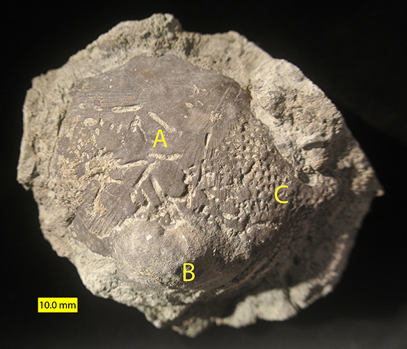
Half borings 152a1Above is a well-used brachiopod from the Upper Ordovician of northern Kentucky (C/W-152; Petersburg-Bullittsville Road, Boone County; Bellevue Member of the Grant Lake Formation). It experienced several events on the ancient seafloor during its short time of exposure. Let’s put a few labels on it and discuss:

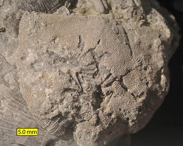
Half borings 152a2Our main topic will be those strange ditch-like borings (A) cut across into the exterior of this brachiopod shell. This is an example of bioerosion, or the removal of hard substrate (the calcitic shell in this case) by organisms. These structures were likely created by worm-like filter-feeders. The shell also has a nice trepostome bryozoan (B) encrusting it (and partially overlapping the borings) and the heliolitid coral Protaraea richmondensis (C), which is distinguished by tiny star-like corallites. The borings are what we need to make sense of in this tableau. Here’s another set on another brachiopod:

Half borings 152bThis closer view of a brachiopod shell exterior from the same locality shows two of these horizontal borings. The mystery is why we see only half of the boring. These are apparently cylindrical borings of the Trypanites variety, but they should be enclosed on all sides as tubes. Why is half missing? It is as if the roofs have been removed. I think that is just what happened.

Half borings 152cThis encrusted and bored brachiopod, again from the same locality, gives us clues as to what likely happened. Here we see an encrusting bryozoan and those borings together. The borings cut through the bryozoan down into the brachiopod shell. Could it be that encrusting bryozoans provided the other half of the borings?

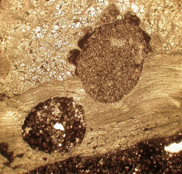
BoringXsectHere’s a test of that idea. Above is a cross-section through the boundary between an encrusting bryozoan (above) and a brachiopod shell (below). It was made by cutting through the specimen, polishing it, and then making an acetate peel. The bryozoan shows the modular nature of its colonial skeleton, and the brachiopod displays its laminar shell structure. The two round features are sediment-filled borings running perpendicular to the plane of the section. The boring on the left is completely within the brachiopod shell; the one on the right is cut along the interface of the bryozoan and brachioopod. Remove the bryozoan and we would have a half-boring as discussed above.

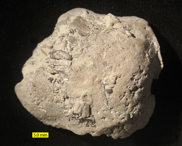
Half borings 152eIf that postulate is true, it means that the encrusting byozoans must have been removed from the brachiopod shells, taking the other halves of the borings with them. We should thus find bryozoans that “popped” off the shells with the equivalent half-borings on their undersides. You know where this is going. The bryozoan above (same locality) shows its upper surface. Note that there are a scattering of tiny borings punched into it.

Half borings 152fThis is the underside of the bryozoan. We are looking at its flat attachment surface. It was fixed to a shell of some kind (I can’t tell what type) and became detached from it. You see the half-borings along with vertical borings drilled parallel to the attachment surface. It appears that small organisms drilled into the bryozoan zoarium (colonial skeleton) on its upper surface, penetrated down to the boundary with the brachiopod shell, and then turned 90° and excavated along the boundary between brachiopod and bryozoan. This makes sense if they were creating a dwelling tube (Domichnia) that they would want surrounded by shell. Punching straight through the bryozoan and brachiopod would leave them in a tube without a base. What would this look like from the inside of the brachiopod shell?

Half borings 152dThis time we’re looking at the interior of a brachiopod shell (same location) that has been exfoliated (some shell layers have been removed). The horizontal borings are visible running parallel to the shell.

Horizontal in bivalveThis view of an encrusted bivalve shell may help with the concept. In the top half you see an encrusting bryozoan. In the bottom you see bivalve shell exposed where the bryozoan has been broken away. Cutting into that shell are the horizontal borings. Their “roofs” were in the now-missing parts of the bryozoan.

There are two conclusions from this hypothesis: (1) There was a group of borers who drilled to this interface between bryozoan and brachiopod skeleton, detected the difference in skeleton type, and then drilled horizontally to maintain the integrity of their tubes; (2) the bryozoans were cemented to the brachiopods firmly enough that the borers could mine along the interface, but later some bryozoan encrusters were removed, leaving no trace of their attachment save the half-bored brachiopod shell. This latter conclusion is disturbing. A tacit assumption of workers on the sclerobionts (hard-substrate dwellers) of brachiopods and other calcitic skeletons is that the calcitic bryozoans cemented onto them so firmly that they could not be dislodged. We could thus record how many shells are encrusted and not encrusted to derive paleoecological data about exposure time, shell orientations and the like. But if the robust bryozoans could just come off, maybe that data must be treated with more caution? After all, bryozoans that were removed from unbored brachiopods could leave no trace at all of their former residence.

Two students and I presented these ideas at a Geological Society of America meeting eight years ago (Wilson et al., 2006), but we never returned to the questions for a full study. Now a new generation of students and I have started a project on this particular phenomenon of sclerobiology. It will involve collecting more examples and carefully dissecting them to plot out the relationship between the borings and their skeletal substrates. We also want to assess the impact these observations may have on encruster studies. Watch this space a year from now!

References:

Brett, C.E., Smrecak, T., Hubbard, K.P. and Walker, S. 2012. Marine sclerobiofacies: Encrusting and endolithic communities on shells through time and space, p. 129-157. In: Talent, J.A. (ed.), Earth and Life; Springer Netherlands.

Smrecak, T.A. and Brett, C.E. 2008. Discerning patterns in epibiont distribution across a Late Ordovician (Cincinnatian) depth gradient. Geological Society of America Abstracts with Programs 40:18.

Wilson, M.A., Dennison-Budak, C.W. and Bowen, J.C. 2006. Half-borings and missing encrusters on brachiopods in the Upper Ordovician: Implications for the paleoecological analysis of sclerobionts. Geological Society of America Abstracts with Programs 38:514.

Posted in Uncategorized | Tagged , , , | 4 Comments

Wooster’s Fossils of the Week: Trace fossils making ghostly shells (Upper Cretaceous of Mississippi)

Entobia gastropod Prairie Bluff Chalk FormationThe unusual fossil above was collected by Megan Innis (’11) and myself in Mississippi during a May 2010 paleontological expedition with Caroline Sogot and Paul Taylor of The Natural History Museum, London. That splendid trip has contributed already to one high profile publication (Sogot et al., 2013) and no doubt more will come from the excellent collections we made. All the fossils in this post came from the Prairie Bluff Chalk Formation (Maastrichtian) exposed at the intersection between Highway 25 and Reed Road in Starkville, Mississippi (locality C/W-395).

The specimen is a marine gastropod (fancy name for a snail), which is hard to believe considering no shell is preserved. The shape of the original aragonitic shell has been taken by a series of interlocking blobs, each with a sediment-filled tube extending outwards. These are casts of chambers made by a boring clionaid sponge. The resulting trace fossil is known as Entobia, a form we have seen several times in this blog. The sequence of events: (1) The sponges excavated cavities connected by tunnels into the aragonite shell of the gastropod, maintaining connections to the seawater for filter-feeding; (2) the cavities and tubes filled with fine-grained calcareous sediment after the death of the sponges; (3) the aragonite gastropod shell dissolved away, probably at the same time the sediment filling the cavities was cemented; (4) the fossil was exhumed as a series of natural casts of the sponge cavities — the trace fossil Entobia.
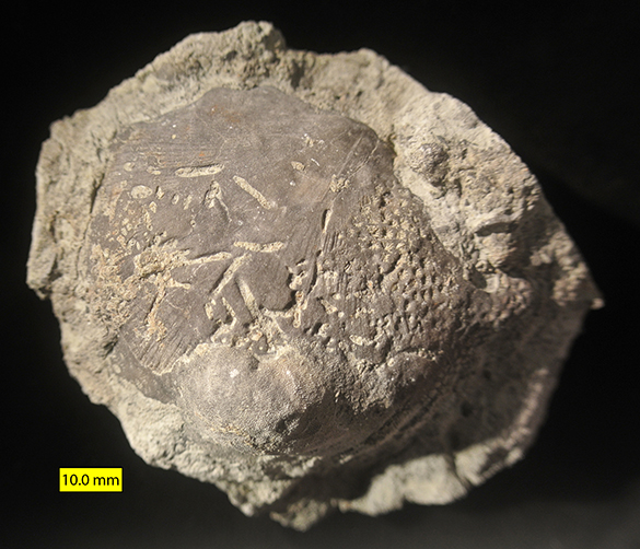
Entobia bivalve 1 exterior Prairie Bluff Chalk FormationThere were many other such fossil ghosts at this locality, such as the apparent bivalve shell fragment above.
Entobia cast close Prairie Bluff Chalk FormationIn this closer view (taken with my new extension tubes on the camera) we see some of the interlocking sponge chamber casts. On the surfaces of some you can just make out a reticulate pattern that represents tiny scoop-like excavations by the sponges. In the upwards-extending tubes there are a few green grains of the marine mineral glauconite.

As a paleontologist it is always sobering to see a fossil preserved in such an odd way. Were it not for these circumstances of boring, filling and cementation, the shells would have completely disappeared from the fossil record. Every fossil we have, really, is a victory of improbable preservation.

References:

Bromley, R.G. 1970. Borings as trace fossils and Entobia cretacea Portlock, as an example. Geological Journal, Special Issue 3: 49–90.

Schönberg, C.H. and Shields, G. 2008. Micro-computed tomography for studies on Entobia: transparent substrate versus modern technology, p. 147-164. In: Current Developments in Bioerosion. Springer; Berlin, Heidelberg.

Sogot, C.E., Harper, E.M. and Taylor, P.D. 2013. Biogeographical and ecological patterns in bryozoans across the Cretaceous-Paleogene boundary: Implications for the phytoplankton collapse hypothesis. Geology 41: 631-634.

Sohl, N.F. 1960. Archeogastropoda, Mesogastropoda, and stratigraphy of the Ripley, Owl Creek, and Prairie Bluff Formations, p. A1-A151. In: Late Cretaceous gastropods in Tennessee and Mississippi: U.S. Geological Survey Professional Paper 331-A.

Taylor, P.D. and Wilson, M.A. 2003. Palaeoecology and evolution of marine hard substrate communities. Earth-Science Reviews 62: 1-103.

Wilson, M.A. 2007. Macroborings and the evolution of bioerosion, p. 356-367. In: Miller, W. III (ed.), Trace Fossils: Concepts, Problems, Prospects. Elsevier, Amsterdam, 611 pages.

Posted in Uncategorized | Tagged , , , | Leave a comment