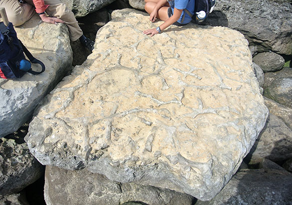
I collected this lump of a specimen during my dissertation research in the Bird Spring Formation (Carboniferous-Permian) of southern Nevada. It was found in a richly-fossiliferous Upper Carboniferous (Moscovian) portion near Mountain Springs Pass, which is about 40 km southwest of Las Vegas. It is a chaetetid, which at the time I interpreted conventionally as a singular extinct sponge in the genus “Chaetetes“. Since then we’ve learned a lot more about chaetetids. (And about the stratigraphy of the Bird Spring Formation. I wish we had sequence stratigraphy way back then!)
 Excellent and thorough work, especially by Ron West, has shown that the chaetetids are “hyper-calcified” members of the Class Demospongiae of the Phylum Porifera. They are sponges indeed, but the tubular chaetetid skeleton is found in at least three orders of the demosponges, including living ones. The chaetetid skeleton, which consists of very thin tubes (as shown above) is polyphyletic, meaning several groups of organisms converged on the same form.
 In this oblique section of a chaetetid you can see the calcitic tubules, somewhat blurred by recrystallization.
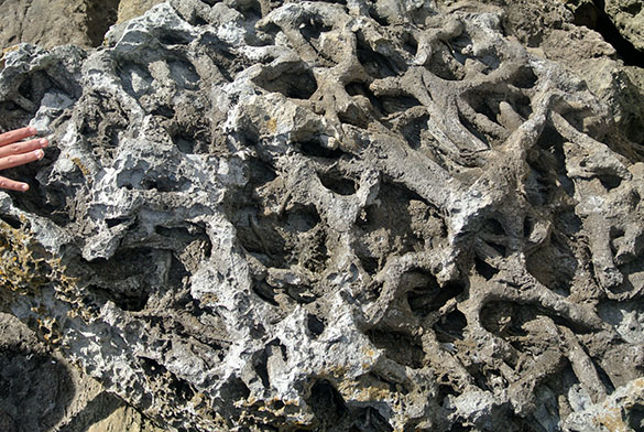
 Here is a cross-section through one of the Bird Spring chaetetids. The tubules are very thin and long, somewhat resembling hair. Chaeto– comes from the Greek chaite for “hair or hairy”.
Now we know from systematic studies that the fossil “chaetetids” cannot be classified from their tubular skeletons alone. Without evidence of the spicules (which are rarely found, or at least recognized) and original mineralogy of the skeleton (many are recrystallized or, like the one at the top of this entry, replaced with silica) we can only refer to skeletal specimens such as ours as “chaetetid hyper-calcified demosponges”.
This is enough, though, for me to reintroduce them into my Invertebrate Paleontology classes. I had removed them from the teaching collections several years ago because of the confusion as to their status. Now they are at least demosponges, hyper-calcified at that.
References:
Almazán, E., Buitrón, B., Gómez-Espinosa, C. and Daniel Vachard. 2007. Moscovian chaetetid (boundstone) mounds in Sonora, Mexico. In: Vennin, E., Aretz, M., Boulvain, F. and Munnecke, A., eds., Facies from Palaeozoic reefs and bioaccumulations. Mémoires du Muséum national d’Histoire naturelle 195: 269–271.
Martin, L.G., Montañez, I.P. and Bishop, J.W. 2012. A paleotropical carbonate-dominated archive of Carboniferous icehouse dynamics, Bird Spring Fm., southern Great Basin, USA. Palaeogeography, Palaeoclimatology, Palaeoecology 329: 64-82.
West, R.R. 1994. Species in coralline demosponges: Chaetetida. In: Oekentorp-Küster, P., ed., Proceedings of the VI International Symposium on Fossil Cnidaria and Porifera, Munster Cnidarian Symposium, v. 2. Courier Forschungsinstitut Senckenberg 172: 399–409.
West, R.R. 2011a. Part E, Revised, Volume 4, Chapter 2A: Introduction to the fossil hypercalcified chaetetid-type Porifera (Demospongiae). Treatise Online 20: 1–79.
West, R.R. 2011b. Part E, Revised, Volume 4, Chapter 2C: Classification of the fossil and living hypercalcified chaetetid-type Porifera (Demospongiae). Treatise Online 22: 1–24.
West, R.R. 2012c. Part E, Revised, Volume 4, Chapter 2D: Evolution of the hypercalcified chaetetid-type Porifera (Demospongiae). Treatise Online 35: 1–26.
Wilson, M.A. 1985. Conodont biostratigraphy and paleoenvironments at the Mississippian-Pennsylvanian boundary (Carboniferous: Namurian) in the Spring Mountains of southern Nevada. Newsletters on Stratigraphy 14: 69-80.


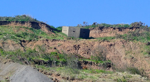
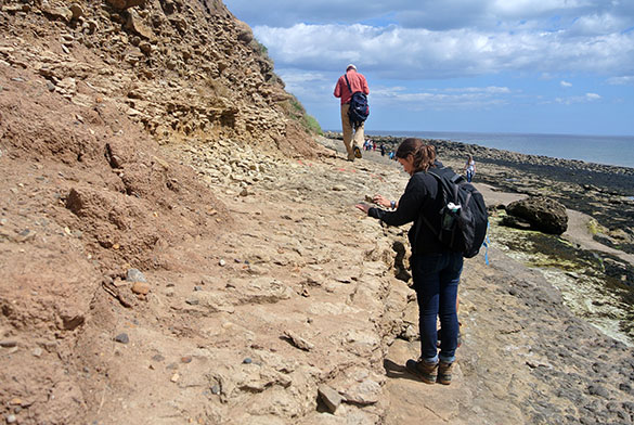
 SCARBOROUGH, ENGLAND (June 8, 2015) — We see many of these World War II concrete defenses along the Yorkshire coastline. This is a pillbox that was likely constructed in 1940 to defend the realm from the Germans. Of course, it was not placed on the sandy beach but up on the steep slopes overlooking the shore. Erosion of that headland since 1940 was complete, leaving this structure on the open beach.
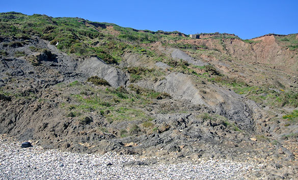
 It is a dilemma, building on these sea cliffs of the northern Yorkshire coast. The substrate here is a “boulder clay”, a Pleistocene glacial deposit known as a diamictite. It is easy to excavate, but flows readily under weight and when wet. The sea hammers away at the foot of these soft cliffs as their tops slump downwards. The heavy concrete gun emplacements and observation posts serve their purpose for a few years, and then eventually fall into the sea as the coast retreats. Seventy-five years of coastal erosion has removed a great deal of the cliffs.


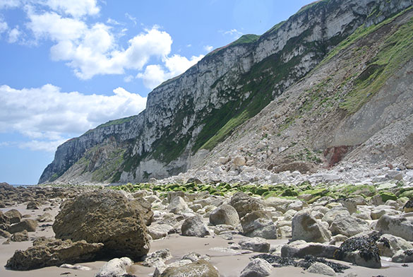
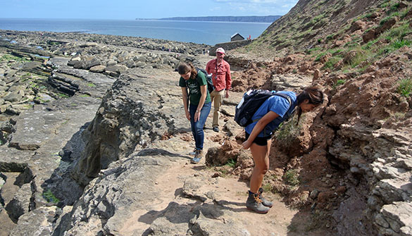
 Given what you’ve seen above, would you buy this seaside house?