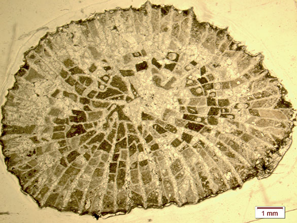
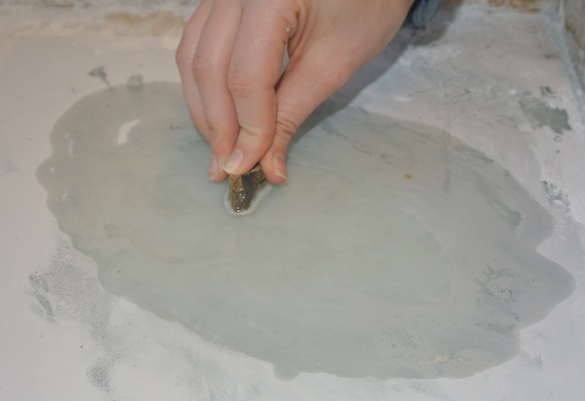
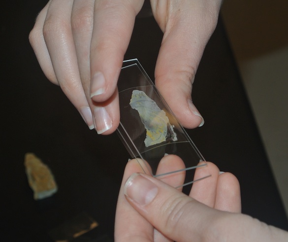
This week’s fossil is again in honor of Annette Hilton (’17), now retired as my Sophomore Research Assistant this year. She has been assessing with great skill a large and diverse collection of scleractinian corals from the Matmor Formation in Hamakhtesh Hagadol in the Negev Desert of southern Israel. These specimens were collected during paleoecological studies by the Wooster paleontology lab and our Israeli colleagues. Above is a fantastic specimen of Microsolena Lamouroux 1821. We are looking at the top of a gumdrop-shaped corallum, with the corallites (which held the polyps) as shallow pits with radiating septa.
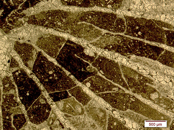
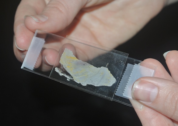
 This is the reverse view of the coral, showing its concentric growth lines. The jumble in the center is shelly debris on which the coral originally established itself on the muddy seafloor. Note that the preservation here includes numerous little circles. These are centers of silica replacement called beekite rings (a form of chalcedony).
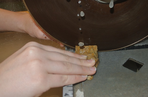
 Above is a closer view of the beekite preservation. The silica circles have concentric rings of their own. This kind of preservation (a type of silicification) is common in the Matmor Formation. Corals like this one started as aragonitic skeletons and then were replaced by calcite and silica. I suspect the calcitization took place first because of the fine degree of preservation for most of the corallum.
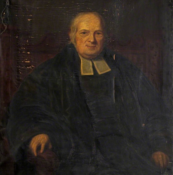 So how do we get a term like beekite? Meet Rev. Henry Beeke (1751-1837), a rather unlikely scholar to make it to this blog. Beeke was an Oxford graduate in divinity, with a strong concentration in history. He became a prestigious Regius Professor of Modern History at Oxford. He was sought after as an expert of economics and taxation at the beginning of the 19th Century. As with many divines of the time, Beeke also pursued natural history, specializing in botany. At some point in his career he was noted as having described what we now call beekite, but I can’t find where in the literature. All I have is a brief mention of “Beekite, a new mineral, named after Dr. Beeke, at the Corbors” in Blewitt (1832, p. 15). Henry Beeke had the distinction of seeing this mineral variety named after him during his lifetime, which is rare in science.
References:
Blewitt, O. 1832. The panorama of Torquay: a descriptive and historical sketch of the district comprised between the Dart and Teign. Second edition. London: Simpkin and Marshall, 289 pages.
Church, A.H. 1862. XV. On the composition, structure, and formation of Beekite. The London, Edinburgh, and Dublin Philosophical Magazine and Journal of Science 23(152): 95-103.
Nose, M. and Leinfelder, R. 1997. Upper Jurassic coral communities within siliciclastic settings (Lusitanian Basin, Portugal): implications for symbiotic and nutrient strategies. Proceedings of the 8th International Coral Reef Symposium 2: 1755-1760.
Pandey, D.K., Ahmad, F. and Fürsich, F.T. 2000. Middle Jurassic scleractinian corals from northwestern Jordan. Beringeria 27: 3-29.
Pandey, D.K. and Fürsich, F.T. 2001. Environmental distribution of scleractinian corals in the Jurassic of Kachchh, western India. Journal Geological Society of India 57: 479-495.
Pandey, D.K. and Fürsich, F.T. 2005. Jurassic corals from southern Tunisia. Zitteliana 45: 3-34.
Wilson, M.A., Feldman, H.R., Bowen, J.C. and Avni, Y. 2008. A new equatorial, very shallow marine sclerozoan fauna from the Middle Jurassic (late Callovian) of southern Israel. Palaeogeography, Palaeoclimatology, Palaeoecology 263: 24-29.
Wilson, M.A., Feldman, H.R. and Krivicich, E.B. 2010. Bioerosion in an equatorial Middle Jurassic coral–sponge reef community (Callovian, Matmor Formation, southern Israel). Palaeogeography, Palaeoclimatology, Palaeoecology 289: 93-101.