Wolf Lake and the Surrounding Landscape, Glacier Bay, Alaska

Members of the Wooster Tree Ring Lab had a great opportunity to travel to a seldom-visited part of Glacier Bay National Park and Preserve – a transect from Wolf Lake to Burroughs Glacier. We were there because there is 2500 year-old forest remnant that was overrun by ice. The ice has gone and continues to melt. Our interest is recovering these logs is to fill a gap millennial-scale tree-ring record from the Gulf of Alaska. The recently exposed logs are being lost to science each year as they flush out into the sea and rot away in this hypermaritime climate. Wooster student independent studies (ISs) in the region quite literally have surrounded this Wolf Lake site with their research, and over the last 10 years we have honed into this key location from all directions.

A view of Mount Wright through a gap in drift and bedrock. Tree-ring records from the flanks of Mt. Wright were part of a study led by Stephanie Jarvis and sampled by Sarah Appleton, who did their thesis work in the region.

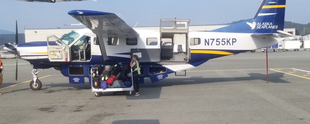
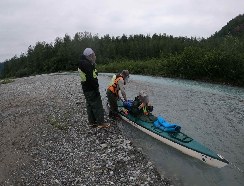
We flew into the site this year. In previous years we attempted to walk in twice and once we were successful. I recommend the flying in – the brush and terrain makes it a brutal walk from Muir Inlet. Previous students Willy Nelson, Zach Downes, Dan Misinay, Jeff Gunderson, Andrew Wayrynen and  Jesse Wiles were some of those that would have appreciated a float plane ride into the lake.

Nick Wiesenberg at the head of a fan on Minnesota Ridge – in the distance is the blue of Wolf Lake and beyond is Muir Inlet. The ice covered this entire scene 100 years ago.


The flight over from Juneau afforded excellent views of Casement Glacier where students Sarah Appleton and Joe Wilch worked. 

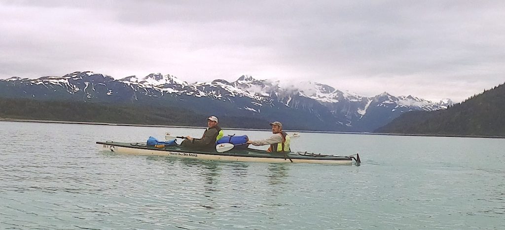
Below is the sediment-charged plume from Casement Glacier as the Casement River empties out into Adams Inlet. IS students Jenn Horton and Lauren Vargo and I paddled through these waters on our way into the Inlet a some years ago.


Back to the Wolf Lake Basin – the upper reaches of the river and the pass (Glacier Pass) that we used to get to the Burroughs Glacier Basin.

We were there for the logs and here Nick is sneaking up on a potential sample.

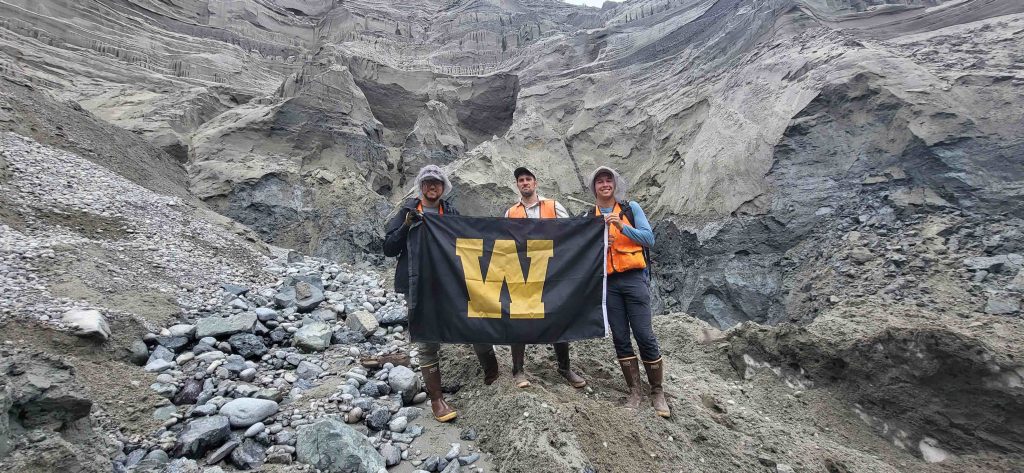
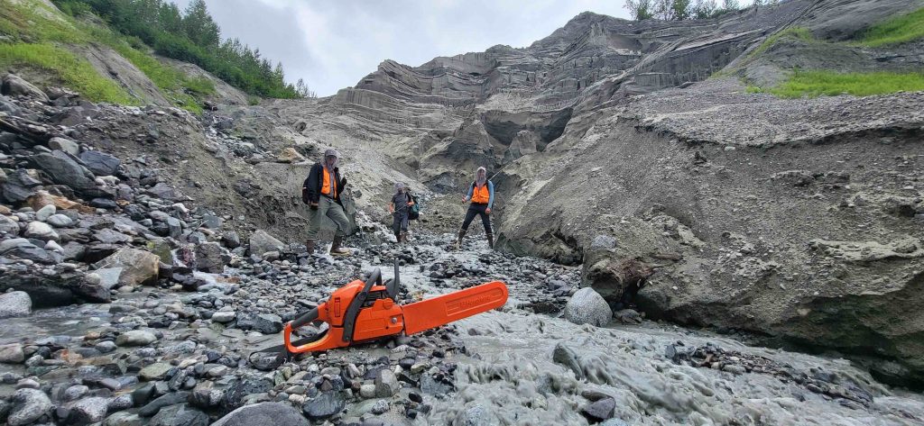
The logs were dispersed in the river as well as hiding throughout some of the coarsest and most angular fans that ever existed. One can appreciate the weathering that is taking place on these fresh surfaces in this hypermaritime climate. The dot in the middle of the fan is Nick. 

The fans were more like rock glaciers and frankly we do not really understand these systems that are less than 100 years old.

I have to give credit to Nick for his tenacity and drive in systematically covering these fan surfaces and sampling the best hemlock logs we could find. This maps shows the sample sites – they are disperse across the landscape, but the logs are there and they cannot hide for long.

Nick coring a log beneath a snow bridge at the head of a fan.

The samples in the upper right portion of the map are taken from the flanks of the remnant Burroughs Glacier (named after the NY naturalist John Burroughs) who was a friend of John Muir and a member of the Harriman Expediti0n. At least five dissertations were written by glacial sedimentologists who studied ice-contact deposition along the margins of Burroughs – they include researchers from The Ohio State University, the University of Wisconsin – Madison and Michigan State. Wooster students Sarah Appleton, Andy Nash and Abby Vanlueven all worked in the area just south of Burroughs.

 Nick standing on the dead ice of Burroughs Glacier. Below he takes a core from a unsuspecting log.


    The granite in the area preserves dome nice glacial features – here a bullet-shaped boulder complete with a striated surface and a plucked (right side) end.

Must be some kind of bits of host rock (dark clasts) incorporated in a magma. 


We are lucky to be collaborating on this project with others on this project from the University of Alaska – Fairbanks and the Park Service. Dr. Ben Gaglioti worked earlier in the summer at another location along Glacier Bay’s wild outer coast – his work was written up in a 3-part series by Alaska Science writer Ned Rozell in the Juneau Empire, here, here and here. This project was funded by the National Science Foundation Paleoclimate Program (Awards: P2C2-2002561 and 2002454).

Posted in Uncategorized | Tagged , | 4 Comments

Apple Creek & Trout Unlimited

Guest bloggers: Chamari Abercrombie and Ellen Yoon.

Good evening Wooster community, today the AMRE Water Team visited Apple Creek with Trout Unlimited to analyze the water quality of Apple Creek. To do so, we looked at macroinvertebrates and collected data from the transducer, which measures water level that we placed in the stream a few weeks ago. Special thanks to Dr. Skip Nault for teaching us about the method and what it indicates about water quality.

Grace Hodges before she goes in to use the dip net method to collect macroinvertebrates.

  • Chamari Abercrombie and Layali Banna used the kick seine method to collect macroinvertebrates. Next, Chamari used water to wash the macroinvertebrates into the silver pan while Layali held the kick seine at an angle to prevent the loss of macroinvertebrates.

  • Dragonfly larvae and Damselfly larvae were collected during the dip net method.

  • Overall data for the collection of macroinvertebrates and water quality – the indicators suggest excellent water quality. – The END. We hope you enjoyed our content.
Posted in Uncategorized | Tagged | 3 Comments

Wooster fieldwork resumes at Brown’s Lake Bog on a gorgeous day

Wayne County, Ohio — It was a perfect day for Wooster Geologists to do some aquatic fieldwork. It was my first day of fieldwork since March 2020 in Utah. This time I wasn’t doing much actual work, though — I watched our geological technician Nick Wiesenberg and Professor Greg Wiles apply their considerable field skills and took these photographs.

We drove to Brown’s Lake Bog in southern Wayne County to collect sediment and plankton samples for Justine Paul Berina’s Senior Independent Study project on diatoms as well as for other grant-supported studies. Nick is shown in the top photo heroically casting a plankton net into the bog to sample microscopic organisms in the top of the water column. We hope we find lots of living diatoms. We know there will be lots of floating algae!

Brown’s Lake Bog is state property operated as a nature preserve by The Nature Conservancy. Wooster has long-standing permission to do scientific work there. It is a fantastic natural laboratory.

Nick is preparing the plankton net for casting. Greg is holding a clear plastic tube we will use to collect a sediment core.

The net is now tied to the platform after use so most of the water can drain through. It took awhile because there was so much suspended algae.

Now Nick and Greg are preparing the core sampling device. The white part above the clear tube has a valve that allows water to pass through when the tube is thrust into the sediment. It closes when the tube is removed, essentially sucking up the enclosed sediment.

Off Nick and Greg go across the floating sphagnum mat to the water’s edge.

Nick is now plunging the sediment collector into the muck.

I’d like to call this the Reverse Iwo Jima maneuver as Greg and Nick remove the sediment-filled core.

The tube is now filled with sediment and water. Success!

Our aquatic scientists are now carrying the precious samples and equipment back to our vehicles. Well done.

Watch this blog for our data and observations from this work in a few months!

Posted in Uncategorized | Tagged , | 2 Comments

Laboratory microphotography in the Department of Earth Sciences at The College of Wooster (Part 2)

This is the third in a series on laboratory photography in the Department of Earth Sciences at Wooster. In a comment on a Fossil of the Week post last month, Wooster Geologist Alumnus Dr. Bill Reinthal asked if I could describe how we make our blog and other photographic images. I started last week with a post on our macrophotography equipment and techniques. You may want to read that post first for our general photographic processes. Next we had a post on our microphotography using a dissecting microscope with reflected light. This last post is on our microphotography through a petrographic microscope using transmitted light. As I cautioned before, I am not a professional photographer, and my departmental colleagues do plenty of their own excellent photography.

Above is an image of a thin-section cut from an oolitic limestone, the Middle Jurassic Carmel Formation of southwestern Utah. (You may see a theme here! I’ve used the same rocks and fossils in this three-part series to compare the various photographic techniques.) This photograph was taken with the equipment described below. I added the scale later with Adobe Photoshop.

This is our petrographic microphotography station in Dr. Meagen Pollock‘s petrology lab. On the left is a Mac computer running the imaging software from Lumenera (Infinity). Again our wonderful Geological Technician Nick Wiesenberg has written a detailed, easy-to-follow guide to using the system. On the right is the petrographic microscope with a thin-section on the stage.

Another view of the arrangement.

The camera is an Infinity 5 (5.1 Megapixel). It makes fantastic images.

Here again is that Carmel Formation oolitic limestone, this time seen as an acetate peel. Note the differences between the thin-section (top image of this post) and the peel of the same rock. They both show unique details. This is why I like to make both peels and thin-sections of carbonate rocks I’m studying.

Layali Banna (’22) took this microphotograph so we could get some hard-rock, cross-polar action in these blogposts. Feldspars! The scale bar is an example of what the system provides.

Here’s another cross-polar microphotograph from Layali. I don’t know the rock, but it looks like a micaceous sandstone. Thanks, Layali!

These three entries on our laboratory photography systems (macrophotography, microphotography part 1, microphotography part 2) are designed to show our current and future students what we can do in our department. Who knows how long this blog will survive in cyberspace, but maybe someday future Wooster Geologists will arrive here and marvel at our Old Ways!

Posted in Uncategorized | Tagged , | 2 Comments

Laboratory microphotography in the Department of Earth Sciences at The College of Wooster (Part 1)

In a comment on a Fossil of the Week post last month, Wooster Geologist Alumnus Dr. Bill Reinthal asked if I could describe how we do our lab photography in the Earth Sciences department. I started what will be a three-part series last week with a post on our macrophotography equipment and techniques. You may want to read that post first for our general photographic processes. Today I’m showing our photographic system that uses a dissecting microscope with reflected light. Later we’ll have a post on our microphotography through a petrographic microscope using transmitted light. As I wrote last time, I am not a professional photographer, and my other departmental colleagues do plenty of their own photography. I’m the one who tends to write the most blog posts! (This is entry #1109 for me …)

The above image is of a limestone bedding plane from the Carmel Formation (Middle Jurassic) of southwestern Utah. You can see most of the grains are carbonate ooids, with a scattering of crinoid (Isocrinus nicoleti) debris, including a beautiful star-shaped columnal. (The high-resolution version can be found here.) This image was made with the equipment described below. The scale bar was added later using Adobe Photoshop.

This is our dissecting microscope photographic system. On the left is a fiber-optic gooseneck lamp with two light tubes. The tube on the left is closer to the specimen to produce a dominant light source from the upper left (a paleontological convention to ensure uniform shading). The fiber-optic tube on the right is farther away from the specimen so that it provides a softer fill-in light to brighten the shadowed areas. These are easily moved for various lighting effects depending on the specimen. The microscope in the middle is a Nikon SMZ 1500 with a Lumenera Infinity 3 camera attached in the upper right. The camera taps into the microscope’s light path, so you don’t need to use the eyepieces. The Mac computer on the right is running the Lumenera Infinity photographic software. Our ace geological technician, Nick Weisenberg, has written detailed instructions for using the software with ease.

The Nikon SMZ 1500 microscope has a built-in aperture, which should be closed down as far as possible to get the best depth-of-field.

The camera works very well. We also have versions 4 and 5 attached to petrographic microscopes.

That’s it for this dissecting microscope photographic system! The images it makes are excellent. We spend most of our time composing the scenes and constructing the scale bars. Two more example photographs are below.

This is olivine sand from a Hawaiian beach. (The original is here.)

This photographic system is used often by Dr. Greg Wiles and his tree-ring lab students. It works especially well with sanded tree-ring cores, which are essentially two-dimensional. Layali Banna (’22) made this evocative image last week.

These three entries on our laboratory photography systems (macrophotography, microphotography part 1, microphotography part 2) are designed to show our current and future students what we can do in our department.

Posted in Uncategorized | Tagged , , | 2 Comments

Laboratory macrophotography in the Department of Earth Sciences at The College of Wooster

In a comment on a Fossil of the Week post last month, Wooster Geologist Alumnus Dr. Bill Reinthal suggested I describe the processes I use to create images of rocks and fossils for this blog, publications and other outlets. This is a good idea because I can produce an outline of our macrophotography techniques for students and others to use as a reference. My only caveat is that I’m not a professional photographer by any means — I’m a scientist who does a lot of photography, trying to retain whatever useful ideas I’ve stumbled across. Like Bill, I’m still building on skills we learned in the Junior Independent Study Geologic Methods course during the last century! I also want to point out that all our departmental faculty and staff do photography and do it well. The following techniques are from my personal perspectives and experiences.

The image above is typical of my lab photography. It is a limestone bedding plane showing trace fossils in convex hyporelief (thus the sole of the bed; the bed is positioned upside-down). This rock is from the Eagle Mountain Ranch exposure of the Middle Jurassic Carmel Formation in southwest Utah, collected during our epic, star-crossed March 2020 expedition. Note the deep black background that provides the ultimate contrast with the edges of the slab. The rock appears to be floating in space with no distractions around it. The dominant light is coming from the upper left, following paleontological tradition for consistency. The yellow scale was added to the image later “in post” as the pros say.

This is the copy stand arranged to take the top image, minus the camera. I believe the four large lights go back at least to the 1970s, and the platform that holds the camera dates to before World War II! The components — A indicates the four main light bulbs (daylight color temperatures); only the back left one is on to produce the required upper-left dominant light source. B is a fiber-optic gooseneck spotlight pair; it is used with smaller specimens so is off. C is a vertical pole that holds the camera mount; the mount can be clamped anywhere along it. D is the camera mount, which has a wingnut-and-spring system to move it vertically in fine increments. (Tighten that wingnut well or the springs can snap the camera up into your face!) E is the copy stand board covered with black velvet to produce the beautiful light-absorbing background. The essential scale cards are always nearby. F is a white piece of cardboard with small light to fill in deep shadows opposite the main lit parts of the specimen. Usually reflection alone is enough fill light, but sometimes I need an extra boost.

Another angle on the copy stand system. You can better see the cantilevered camera mount and the reflective cardboard.

The fancy camera is mounted! This is our latest departmental camera, purchased just last month. It is a Nikon digital single-lens reflex (DSLR) camera, model D5600. The zoom lens attached here is 18-55 mm focal length. I also use a 40 mm lens for really close work. This camera is a dream with its tilting touch screen, superb automatic focus, and multiple shooting controls.

Typically I use the aperture-priority shooting mode, shown by the yellow arrow. This gives me control of the depth-of-field since the shutter speeds can be slow with the sturdy mount. I use “live view”, which allows me to see the composed image on the camera screen. I simply lightly touch the screen where I want the primary focus and the camera takes the picture. Amazing. Occasionally I use exposure adjustments, but most of the time the lighting is good enough as is.

Here is a closer view of the most prominent trace fossil taken with the 18-55 mm lens. By the way, I have no idea what trace fossil this is. Could it be sea anemone burrows? Snail burrows? If you recognize it, let me know!

This image shows as close as I can get with the 40 mm lens, which is closer than any of our previous cameras could do. The rock is an oolite from the same outcrop of the Carmel Formation. Note the crinoid bits scattered amongst the ooids. This is the upper end of the size range for which we do microphotography.

When I’ve finished a photography session I download the images from the memory card into my MacBook Pro computer and do the post-processing with Adobe Photoshop. Most of this work is cropping, with some exposure adjustments and occasional dodging and burning in (terms that only make sense if you remember the days of film and darkroom printing!). The last item I add is the scale bar.

All the image versions on this blog, of course, have been reduced in size for quicker loading on the web. Any images I make that could be useful to others are uploaded in their original dimensions into the Wikipedia system (here is my Wikimedia index page) and designated public domain.

If you have any questions, please ask in the comments, by email or on Facebook! Later I’ll have a similar post on our microphotography systems.

These three entries on our laboratory photography systems (macrophotography, microphotography part 1, microphotography part 2) are designed to show our current and future students what we can do in our department.

Posted in Uncategorized | Tagged , , , | 1 Comment

A diatom study begins at Wooster

This happy Wooster Geologist is Justine Paul Berina (’22). He and I have started a project with diatoms found in mud cores taken from Brown’s Lake and Brown’s Lake Bog by Dr. Greg Wiles, Nick Weisenberg, various crews from the University of Cincinnati, and countless students of Dr. Wiles. Justine and I are honored to join this productive team that has studied this lake and bog system for many years.

Diatoms are microalgae with two-part siliceous skeletons (frustules) found in just about every aquatic system. They manufacture from 20 to 50% of our atmospheric oxygen and cycle billions of tons of silica through the environment. If you want more details on diatoms, check out what I consider the best website ever devoted to a taxonomic group: Diatoms.org.

Diatoms are especially helpful for assessing the health of water systems because they are very sensitive to aquatic geochemistry and ecological changes. This is how Justine and I are using the Brown’s Lake diatom distributions, essentially as paleoecological tools.

Above is Brown’s Lake Bog, familiar to generations of Wooster students and readers of this blog.

Justine devoted his Junior Independent Study project last semester to examining diatoms from a Brown’s Lake Bog core. He used an existing collection of smear slides to first find where diatoms were most common, and then he made his own slides. Justine and I are very fortunate to have the advice of a diatom expert, Dr. Julie Wolin at Cleveland State University. Dr. Wolin corrected our diatom identifications and has given us many ideas for future work.

Our project involves studying the diatoms in core sections before and after agricultural work began in the Brown’s Lake area. We want to see how the lake and bog conditions changed with the anthropogenic modifications of the drainage systems and soils.

This is an image Justine made of our most common Brown’s Lake Bog diatom, Pinnularia.The diatom Stauroneis has a subtle “bowtie” in its center.

There are also numerous siliceous sponge spicules scattered through the cores. We hope they have some paleoecological value as well.

Justine has only a few more days left in the lab before he travels to the University of Delaware for a summer internship. I will continue our work on diatoms until Justine returns in August to begin his Senior Independent Study project on the critters.

Again Justine and I are proud to join the Brown’s Lake crew and look forward to making our contributions to the science!

Posted in Uncategorized | Tagged , , | 3 Comments

Wooster’s Fossils of the Week: Giant Pliocene scallop from Virginia with bonus sclerobionts

Yes, the feature “Wooster’s Fossil of the Week” was retired long ago (all entries still available on this blog), but occasionally I will still cover interesting fossils we come across in the lab or field. The title is now a tradition, even if the items don’t appear every week. No good fossils should be left behind.

The beautiful specimen above was kindly donated to the department last week by Wooster Geologist Alan Troup (’96). He found it and several other specimens in the Yorktown Formation (the Pliocene part) along the York River in Virginia. It will be used in our Paleoecology course next year. These fossil scallops are incredibly abundant, and this is an especially nice one with its numerous sclerobionts (hard-substrate-dwelling organisms). The main shell is Chesapecten jeffersonius, one of the largest scallops ever and the state fossil of the Commonwealth of Virginia. (I don’t know if “Commonwealth Fossil” is a thing.) It is encrusted on the outside only by large barnacles. Near the hinge are numerous perforations from clionaid sponges, making the trace fossil Entobia. It makes for a sweet little community.

Here’s a closer view of Entobia. Each hole leads to a tunnel system in the thick scallop shell.

Other scallops in the collection are encrusted by these thin scleractinian corals with radiating septa inside the  corallites.

The arrow points to a predator drill hole (the trace fossil Oichnus) through the scallop shell. It was made by a naticid gastropod.

Alan’s donation also includes oysters like the above. I’m sure you by now see the included trace fossils!

One of the many interesting questions about these sclerobiont-laden scallops is whether they could do their “swimming” while so heavily encrusted on their exteriors. As you can see in this video, modern scallops can swim today with a good load of passengers.

Reference:

Ward, L.W. and Blackwelder, B.W. 1975. Chesapecten, a new genus of Pectinidae (Mollusca: Bivalvia) from the Miocene and Pliocene of eastern North America. U.S. Geological Survey Professional Paper 861, 24 p.


Posted in Uncategorized | Tagged , , | 3 Comments

Milestones for two Wooster Geologists

There was some good news for the College of Wooster Earth Sciences faculty during the otherwise dreary Pandemic Year. The two cheerful Wooster geologists pictured above in the field (today!) reached important points in their professional lives.

Dr. Meagen Pollock was promoted to Full Professor this spring. This is a big deal in the academic world. The criteria are: “The College expects that faculty portfolios for promotion to Professor will demonstrate significant and sustained achievements in the areas of teaching, scholarship, research, and general value to the College after those that led to tenure and/or the rank of Associate Professor.” She’s going to have to update that faculty webpage!

This year Dr. Greg Wiles was elected a Fellow of the Geological Society of America. The requirements: “Society Fellowship is an honor bestowed on the best of our profession by election at the spring GSA Council meeting. GSA members are nominated by existing GSA Fellows in recognition of a sustained record of distinguished contributions to the geosciences and the Geological Society of America through such avenues as publications, applied research, teaching, administration of geological programs, contributing to the public awareness of geology, leadership of professional organizations, and taking on editorial, bibliographic, and library responsibilities.” Greg’s nomination was in two categories: “Publication of the results of geological research” and “Training of geologists”. No more than two categories could be selected — otherwise he would have more!

Congratulations to Meagen and Greg. They are heroes!

Posted in Uncategorized | Tagged , | 3 Comments

Wooster Memorial Park – Now Part of the Old Growth Forest Network

On 20 April 2021 Wooster Memorial Park became part of the Old Growth Forest Network.  The founder and director, Joan Maloof, visited Wooster Memorial Park forest to officially induct the park into the Network. The Wooster Tree Ring Lab cored some of the white oaks in the park to determine their age and to see how they are responding to the increasingly wetter climate of Northeast Ohio. Nathan Kreuter (Biology) and Nick Wiesenberg (Earth Sciences Dept. Technician) found the oldest trees and helped work up the tree-ring data.

Joan Maloof with the largest hemlock in the park.

Nathan Kreuter cores one of the oldest oaks as part of his tutorial at the Wooster Tree Ring Lab.

The ring-widths of the Wooster Memorial Park chronology. There was a likely time of early logging in the park about 1815 and again in the 1920s
Tree ring widths are most sensitive to April-August total precipitation. The correlation between the ring-widths and precipitation changes over time with the strongest relationship r = 0.6 for the interval 1945-1975. After 1975 the correlation falls off possibly due to the increase in precipitation and loss of sensitivity at the site with the abundant moisture.

Friends of the trees and Friends of Wooster Memorial Park with Joan Maloof.

Posted in Uncategorized | Tagged , | 2 Comments