San Diego, CA – If you’ve been following our adventures, you know that we’ve started a project on Black Mountain with our collaborators at the University of San Diego. We’ve dedicated a significant portion of our time in California to sample preparation, and today we see the results of all of our hard work.
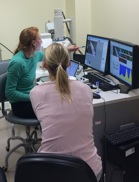
In order to address our research questions, we need to understand the compositions of the minerals, rocks, and soils that are present at the field site. To analyze mineral compositions, we are using a scanning electron microscope equipped with an energy-dispersive detector (SEM-EDS). The electron beam interacts with a polished rock specimen to produce characteristic X-rays. The detector separates those X-rays by energy, correlates the energy to specific elements, and maps the distribution of elements in the sample. This technique allows us to determine the compositions of individual minerals in our rocks.

Elizabeth Johnston (USD graduate student) and Dr. Beth O’Shea (USD) are examining mineral compositions using an SEM-EDS.

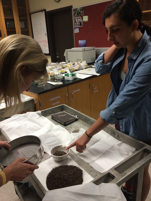
Dr. O’Shea and Amineh AlBashaireh (’18) examine soil samples and discuss analytical strategies.
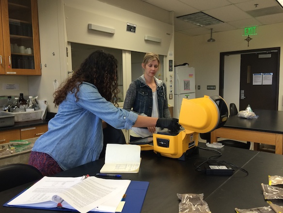
To analyze the bulk compositions of our soil samples, we’re using a benchtop X-ray fluorescence spectrometer (XRF). The XRF uses an X-ray beam to generate X-rays from the samples. The generated X-rays are characteristic of specific elements, which the XRF measures and compares to a calibration curve to calculate a concentration. This XRF model is equipped with several modes for analyzing soil or ore samples and allows us to analyze bulk compositions without destroying the sample.

Amineh is analyzing her soil samples with the benchtop XRF. She will use these data to guide her analytical work when we return to Wooster.


Since soil samples are often so full of clay minerals, do you worry about preferred orientation of them, flattening out preferentially on the surface, when you make pressed plugs for XRF, giving you a higher-than-intended signature for those components of the mixture?
In the old days, we used to worry quite a bit about that with any rocks that had significant layer silicates, but I have no knowledge about how soil samples are handled to avoid that, unless you fuse the samples, rather than pressing them. I suppose you could also randomize the orientation of the particles in the samples by suspending them in an epoxy mix, the way we used to powder samples onto something like Vaseline on a glass slide before doing XRD scans. Just curious!
Great observation! For X-ray Diffraction (XRD), we typically mount clays with a preferred orientation so that we can get a good measurement of the d-spacing between the sheets and thus a more confident identification. For X-ray Fluorescence (XRF), because we’re generating X-rays that are characteristic of the elements that are in the sample, we don’t care as much as preferred orientation. In fact, we actually try to obliterate all of the crystal structure to minimize matrix effects by melting the material and making a glass disk. So, the benchtop XRF was good for a quick compositional measurement, but back at Wooster, we’ll fuse the samples and get a more accurate analysis.