Experiential Learning on Ice (with some water)

Tom Lowell, Aaron Diefendorf and four students from the University of Cincinnati met up with the Wooster Geologists to core Browns Lake. We thank Marvin Sandy, who manages the bog for the Nature Conservancy for guidance and permission to do this work.
coresite

Coring Browns Lake from an ice platform on a winters day in Northeast Ohio. Four cores were taken – the longest of which was 17 meters. The mud in the cores is a record of 15,000 years of environmental change since the last Ice Age.
Browns Lake Bog is own by the Ohio Department of Natural Resources and managed by the Nature Conservancy.

Browns Lake Bog is owned by the Ohio Department of Natural Resources and managed by the Nature Conservancy.

pitcher

Pitcher plants are among the special biology of the bog. Note the ice forming within the pitcher.

auger

First a hole is augured to determine water depth in the basin. It is about 5 feet deep with 4-6 inches of ice.

A look at the drilling rig - anchored in the ice, tied down with ice screws.

A look at the drilling rig – anchored in the ice, tied down with ice screws and straps.


pipe

A look down the long axis of the drill pipe – note the corer sticking out of the ice stored in the lake to prevent it from freezing. The water is the warmest place on the site.

Tom explains the theory and Doug and Nick move into the practice phase.

Tom, the core boss,  explains the theory and Doug and Nick move into the practice phase.

humor

Tom and Michael share a coring joke – it help to have a sense of humor standing on ice for 6 hours at 15 degrees F with a breeze.

meter

Another meter of core is brought up – ready to be described, wrapped and archived. About half of the 40 meters of core went to Cincinnati for further analyses.

Lunch on the boardwalk

Lunch on the boardwalk

oscar_andy

Oscar and Andy take a break from the core archiving. Note the water that moves up through the hole in the ice. The weight of the rig and crew cause elastic and some plastic deformation to occur – after the rig is removed the ice slowly pops back into shape.

The last of the gear moves to the parking lot.

The last of the gear moves to the parking lot.

Thanks to Jesse Wiles for the photography.

The geese point the way back to Wooster. Thanks to Jesse Wiles for the photography.


Posted in Uncategorized | Tagged , | 2 Comments

Wooster’s Fossil of the Week: A crinoid-rich Lower Carboniferous siderite concretion (part III — those crinoids had company)

Crinoid with platyceratid (cross-section) 585The last installment of our analysis of a Lower Carboniferous fossiliferous siderite concretion given to the department by Sam Root. In part I we looked at the crinoid stems and calices on the outside and discuss the formation of siderite concretions and the preservation of this particular assemblage. In part II we had our first look at polished sections of the concretion, taking special note of the crinoid stem morphology and its replacement by the mineral marcasite. For part III you were promised a molluscan surprise.

In the top view you can see that we have a section that fortuitously cut right through the center of a crinoid head. The stem is visible at the bottom, with the calyx and attached arms above. Crowning the calyx is a thin semi-circle of shell nestled open-side-down across the crinoid oral surface. This we can tell from the shell morphology is a parasitic platyceratid gastropod caught in place on its crinoid host. Nice.
Platyceratid Lower Carboniferous 585 annotatedThree years ago we received a fossil donation from the Calhoun family of local Lower Carboniferous fossils, including this beauty pictured above. It is a crinoid calyx (you can tell by the polygonal plates) with a cap-shaped platyceratid gastropod (Palaeocapulus acutirostre) preserved in place on top of it between the arms (now missing). I drew a line across the image to indicate the likely plane of section through a similar pair in our siderite concretion. In section the platyceratid would be recorded as a thin shelly top on the calyx.

Platyceratids have long been known as Paleozoic associates of crinoids. For many years we thought of them as simply coprophagous, meaning they were consuming crinoid feces as they exited the anus. (Awkward conversation, I know.) Careful work by Tom Baumiller (1990) showed that this arrangement (which would not have directly harmed the crinoid because it was, after all, done with the food) was likely not the case. He found trace fossil evidence that the platyceratids were likely accessing crinoid stomach contents directly through some sort of proboscis, and that these parasitized crinoids were stunted in their growth and thus directly harmed (but not killed — no good parasite wants to lose its meal ticket). Our new specimen was thus likely a miserable little crinoid, even if it didn’t have a brain to sort out its feelings.
Stem Calyx 121413As one last view of our crinoids in the concretion, look at the detail in the crinoid stem just below the calyx. The lumen is visible in the center of the stem, as well as the alternating ornaments on the columnals.

This has been a fun specimen to examine. Thanks, Sam!

References:

Baumiller, T.K. 1990. Non-predatory drilling of Mississippian crinoids by platyceratid gastropods. Palaeontology 33: 743-748.

Donovan, S.K., and Webster, G.D. 2013. Platyceratid gastropod infestations of Neoplatycrinus Wanner (Crinoidea) from the Permian of West Timor: speculations on thecal modifications. Proceedings of the Geologists’ Association 124: 988–993.

Posted in Uncategorized | Tagged , , | Leave a comment

Wooster’s Fossil of the Week: A crinoid-rich Lower Carboniferous siderite concretion (part II — the inside story)


1 Cross-section macro 2 121413Last week’s specimen was a Lower Carboniferous fossiliferous siderite concretion from an unknown location, but likely from the Wooster area. It was donated to the department by Emeritus Geology Professor Sam Root. The concretion has beautiful crinoids preserved in it, including several stems of at least two types and three calices (crowns or heads).

I took a chance and cut the concretion with a rock saw if there were interesting features on the inside. There were indeed! In the image above you see at the bottom a cross section through a broken crinoid stem showing the articulated columnals. Above it are sections of crinoid arms (the white and grey spots) each trailing a pair of delicate pinnules (the feeding parts of the arms that carried tube feet). The arms are coming from an intact calyx that is not in the plane of the section.
2 Micro 1 121413In this closer view of the above stem we see the complex anatomy of the crinoid stem. We also see the amazing mineralogy of these specimens in a way we could not from the outside. The light brown matrix is, as we’ve said, the concretion made primarily of siderite (an iron carbonate) and clay. The crinoid columnals, which were originally made of calcite (calcium carbonate), have a silvery metallic material replacing them. This is the iron sulfide mineral marcasite. The white mineral on the inside of the stem on the left is quartz (silicon dioxide). It filled in open spaces inside the stem. To confuse things (nothing is ever easy in this business!) on the right end of the stem marcasite has filled in the cavities instead of quartz.
3 Macro close 121413This view of another stem in cross-section shows a fourth mineral in the system: calcium carbonate. It can be seen as the glassy material in the middle of the structure. It is not the original calcite that made up the columnals. It is instead a later mineral that, like the quartz and marcasite in the previous image, filled in open spaces within the stem. The marcasite, quartz and calcite are thus secondary minerals introduced to the fossil long after its burial. We call these chemical and physical changes to the original mineralogy diagenesis.
4 Fearnhead 2008 Fig 2Since this cross-section view of the crinoid stems is surprisingly complicated, here is a diagram from Fearnhead (2008, figure 2). The top is a crinoid columnal looking at its articulating surface. At the bottom is a cross-section. In our crinoids you can easily make out the lumen as a hollow space running through the center of the stems (filled with marcasite, calcite or quartz). The zygum is that portion of the columnal replaced by marcasite.

Lat week I mentioned that there was a molluscan surprise revealed upon cutting open this concretion. I’ll save that for part III of this series. Same channel next week!

References:

Fearnhead, F.E. 2008. Towards a systematic standard approach to describing fossil crinoids, illustrated by the redescription of a Scottish Silurian Pisocrinus de Koninck. Scripta Geologica 136: 39-61.

Posted in Uncategorized | Tagged , , | 8 Comments

Last official meeting of Wooster Team Israel

Team Israel 2013 011014WOOSTER, OHIO — Above you see Wooster Team Israel 2013 veterans Lizzie Reinthal, Steph Bosch and Oscar Mmari (whom I seem to have caught with his mouth full). Since I’m starting a research leave this semester, we took a last chance to have an evening meeting with pizza, lemon dessert, popcorn and a movie in the warm Wilson living room. It is wintry Ohio outside, but we all have memories of the beautiful Negev:

GoodbyeMakhteshGadol070713a

And what was the movie? You really don’t need to ask, do you?

Lawrence poster

Posted in Uncategorized | Tagged , | Leave a comment

Wooster’s Fossil of the Week: A crinoid-rich Lower Carboniferous siderite concretion (part I)

Cobble Top 121413Last year Wooster emeritus geology professor Sam Root generously donated the above pictured siderite concretion to our paleontology collections. He had received it from a friend who didn’t know where it came from originally so we have no location. The fossils in it, though, show it is Lower Carboniferous in age and could well be from local outcrops of the Cuyahoga Formation. Sam knew this is a cool specimen so he wanted to see what we could make of it.

In the top view we can see crinoid stems running transversely across the surface. Remarkably, two crinoid calices (the arm-bearing crown of the crinoid at the top of the stem) are visible. The larger one is in the lower left. You can see the top of the stem to the farthest left, and then the calyx and attached arms to the right. The second calyx is in the upper right with the arms extending down and towards us. Finding one crinoid calyx with the delicate arms still attached is impressive; finding two in the same slab is a real treat.
Siderite Concretion Carboniferous 585Above is the other side of the concretion. Again a crinoid stem can be seen transverse across the surface. This stem is different from those on the other side, though. It does not have external sculpture, and it is separated into distinct pluricolumnals as if someone sawed through it at regular intervals.
Cobble closer 121413A closer view of the above shows yet another crinoid calyx, this one almost entirely buried in the rock with the arms extending to the surface. The arms have smaller sub-arms (pinnules) still attached. Amazing.

The concretion is made of the mineral siderite (an iron carbonate) that precipitated in fine-grained sediments around the fossils after they were buried. This usually takes place under subsurface anoxic and slightly acidic conditions. The crinoids with all their small and easily-disarticulated parts were buried quickly on the ancient seafloor, probably by a storm-induced pulse of silts and clays. The decay of their soft parts produced hydrogen sulfide gas ad carbon dioxide, triggering the geochemistry that caused the precipitation of siderite around them. The hard concretion that resulted was likely in a matrix of soft shale. The strength of the siderite kept the fossils from being crushed by the weight of sediment above. At some point many millions of years later, the shale eroded away and the concretion was freed to be picked up by some lucky person.

The crinoid stem that is divided into regular increments is interesting on its own. These segments with multiple columnals (the poker chip-like individual elements) are called pluricolumnals. They likely broke at pre-set weaknesses in the connective tissue of the living crinoid, something we see in their living descendants. This may have allowed them to break off their stems (autotomize) when in danger so that the calyx and remaining stem could float away for re-establishment elsewhere.

This concretion is so interesting that I (forgive me, Sam) could not resist cutting it open to see what is inside. The inner view is even more fascinating and will be revealed next week in part II of this story. As a teaser, it involves four minerals and a surprising mollusk!

References:

Baumiller, T.K. and Ausich, W.I. 1992. The broken-stick model as a null hypothesis for crinoid stalk taphonomy and as a guide to the distribution of connective tissue in fossils. Paleobiology 18: 288-298.

Gautier, D.L. 1982. Siderite concretions; indicators of early diagenesis in the Gammon Shale (Cretaceous). Journal of Sedimentary Research 52: 859-871.

Posted in Uncategorized | Tagged , , | 4 Comments

Wooster’s Fossil of the Week: Glyptodon carapace fragment from the Pleistocene

Glyptodon carapace fragment Pleistocene 585This is a tiny bit of a large and fascinating Pleistocene animal from Central and South America. It is Glyptodon, an impressively large mammal with bony armor much like its cousin the armadillo. The above fossil is a fragment of that carapace. Each roundel is called a scute.
Glyptodon carapace side 585This is a side view of the above carapace fragment showing its thickness and layered, bony nature.
Glyptodon ReconstructionThis modern reconstruction of Glyptodon (from Wikipedia with a GNU free documentation license) shows its primary features, including the bony shell (the size and shape of a Volkswagen Beatle, as is often stated) and its characteristically large claws. It belongs to the Superorder Xenarthra, which includes armadillos, sloths and anteaters. I see the resemblance. They could not completely go turtle, as it were, but it could pull its head back enough into the shell that the scutes on the top of the skull would protect it like a cap. They had massive jaws and flat grinding teeth typical of a large herbivore. Its squat skeleton had a variety of features to support the heavy shell, including fused vertebrae and elephant-like short, stout limbs. They went extinct only about 10,000 years ago, possibly having been hunted to oblivion by early Americans. There is even some evidence that people used their empty shells as shelters.
Richard_OwenGlyptodon was formally named as a genus in 1839 by the extraordinary Sir Richard Owen (1804-1892). Owen was a giant of natural history through most of the 19th Century. He is most remembered for inventing the term Dinosauria (“terrible lizards”) and for being on the wrong side of history at the beginning of the Darwinian Revolution. He was apparently ambitious to the point of severity, and very tough on his contemporary scientists. Thomas Henry Huxley, for example, despised Owen for his treatment of his colleagues. Ironically, Huxley did considerable work on further describing Glyptodon in 1865. Owen had vision as well as sharp observational skills. He was a primary force in the eventual establishment of the Natural History Museum in London in 1881. It can be argued that this museum set the high standards of accessibility and research we now expect from all such institutions. Sir Richard Owen is such a large and well known figure I can simply refer you to one of many websites describing Owen’s life and contributions.

This post marks three complete years of Wooster’s Fossil of the Week. That’s 156 posts. You can visit the very first post (about a Devonian tabulate coral) and see how the entries have evolved, so to speak. We still have plenty more fossils to describe!

References:

Gallo, V., Avilla, L.S., Pereira, R.C. and Absolon, B.A. 2013. Distributional patterns of herbivore megamammals during the Late Pleistocene of South America. Anais da Academia Brasileira de Ciências 85(2): 533-546.

Huxley, T.H. 1865. On the osteology of the genus Glyptodon. Philosophical Transactions of the Royal Society of London 155: 31-70.

Oliveira, É.V., Porpino, K.O. and Barreto, A.F. 2010. On the presence of Glyptotherium in the Late Pleistocene of northeastern Brazil, and the status of “Glyptodon” and “Chlamydotherium“. Paleobiogeographic implications. Neues Jahrbuch für Geologie und Paläontologie-Abhandlungen 258(3): 353-363.

Owen, R. 1839. Description of a tooth and part of the skeleton of the Glyptodon, a large quadruped of the edentate order, to which belongs the tessellated bony armor figured by Mr. Clift in his memoir on the remains of the Megatherium, brought to England by Sir Woodbine Parish. FGS Proceedings of the Geological Society of London 3: 108-113.

Posted in Uncategorized | Tagged , , | Leave a comment

Wooster’s Fossils of the Week: Rugose corals from the Upper Ordovician of Ohio

585px-LibertyFormationSlab092313College of Wooster student Willy Nelson spotted and collected up this beautiful Liberty Formation slab on our 2013 Invertebrate Paleontology course field trip to the Upper Ordovician of the Caesar Creek area in southern Ohio. There are many exquisite fossils on this apparent carbonate hardground (a cemented seafloor), the most prominent of which are the four linked circular corallites in the top center. They are of the species Streptelasma divaricans (Nicholson, 1875), shown in more detail below.

Streptelasma divaricans (Nicholson, 1875) 585Streptelasma divaricans is a rugose coral, a prominent order that dominated the Paleozoic coral world from the Ordovician into the Permian. Unlike most rugose corals, it usually is found attached to some hard surface like a shell, rock or hardground. S. divaricans is relatively rare in the Upper Ordovician of the Cincinnati area compared to its free-living cousin Grewingkia canadensis. In its adult form (as seen here) it can have about 60 septa (vertical partitions radiating from the center), alternating from small to large and often with a twist at the center. In life there would have been a tentacle-bearing polyp sitting in each of these septate cups (corallites) catching tiny prey as it passed by in the water currents. We presume that they lived much like modern corals today. S. divaricans was, by the way, an invading species in this Late Ordovician shallow sea community.

Streptelasma divaricans was named as Palaeophyllum divaricans in 1875 by Henry Alleyne Nicholson (1844-1899). We met Dr. Nicholson in an earlier blogpost. Astonishingly, one of our  geology majors in the paleontology course this semester is Brittany Nicholson, a direct descendant. Way cool.
WillyBrachiopodLepidocyclusperlamellosus092313Another nice fossil on Willy’s slab (in the upper right) is the rhynchonellid brachiopod Lepidocyclus perlamellosus, shown closer above.
WillyBivalve092313The curved, indented line in the middle of the slab (shown above) appears to be the outline of a bivalve shell. The original shell was made of aragonite and thus dissolved away very early (possibly even on the seafloor before burial). There is not enough shape remaining to identify it. The twig-like fossil with tiny holes above the scale is, of course, a trepostome bryozoan. You didn’t need me to tell you that!

References:

Elias, R.J. 1983. Middle and Upper Ordovician solitary rugose corals of the Cincinnati Arch region. United States Geological Survey Professional Paper 1066-N: 1-13.

Elias, R.J. 1989. Extinctions and origins of solitary rugose corals, latest Ordovician to earliest Silurian in North America. Fossil Cnidaria 5: 319-326.

Nicholson, H.A. 1875. Description of the corals of the Silurian and Devonian systems. Ohio Geological Survey Report, v. 2, part 2, p. 181-242.

Patzkowsky, M.E. and Holland, S.M. 2007. Diversity partitioning of a Late Ordovician marine biotic invasion: controls on diversity in regional ecosystems. Paleobiology 33: 295-309.

Posted in Uncategorized | Tagged , , , | Leave a comment

Wooster’s Fossil of the Week: A trepostome bryozoan from the Upper Ordovician of northern Kentucky

Heterotrypa Corryville 585First, what U.S. state does this delicious little bryozoan resemble? It’s so close I can even pick out Green Bay. This is Heterotrypa frondosa (d’Orbigny, 1850), a trepostome bryozoan from the Corryville Formation (Upper Ordovician) in Covington, Kentucky. I collected it decades ago while exploring field trip sites for future classes. This zoarium (the name for a bryozoan colony’s skeleton) is flattened like a double-sided leaf, hence the specific name referring to a frond. In the view above you can see a series of evenly spaced bumps across the surface termed monticules. A closer view is below.
Heterotrypa closer 585The monticules are composed of zooecia (the skeletal tubes for the individual bryozoan zooids) with slightly thickened walls standing up above the background of regular zooecia. The hypothesized function of these monticules was to make the filter-feeding of the colony more efficient by utilizing passive flow to produce currents and whisk away excurrents from the lophophores (feeding tentacles) like little chimneys. In 1850, Alcide Charles Victor Marie Dessalines d’Orbigny (French, of course) originally named this species Monticulipora frondosa because of the characteristic bumps.
Boring in Heterotrypa 585If you look closely at the zoarium you will see holes cut into it that are larger than the zooecia. A closer view of one is shown above. These are borings called Trypanites, which have appeared in this blog many times. They were cut by some worm-like organism, possibly a filter-feeding polychaete, that was taking advantage of the bryozoan skeleton to make its own home. It would have extended some sort of filtering apparatus outside of the hole and captured organic particles flowing by. It was a parasite in the sense that it is taking up real estate in the bryozoan skeleton that would have been occupied by feeding zooids. It may not have been feeding on the same organic material, though, as the bryozoan. It may have been consuming a larger size fraction than the bryozoan zooids could handle.

References:

Boardman, R.S. and Utgaard, J. 1966. A revision of the Ordovician bryozoan genera Monticulipora, Peronopora, Heterotrypa, and Dekayia. Journal of Paleontology 40: 1082-1108

d’Orbigny, A. D. 1850. Prodro/ne de Paleontologie stratigraphique universelle des animaux mollusques & rayonnes faisant suite au cours elementaire de Paleontologie et de Geologic stratigraphiques, vol. 2. 427 pp. Masson, Paris.

Erickson, J.M. and Waugh, D.A. 2002. Colony morphologies and missed opportunities during the Cincinnatian (Late Ordovician) bryozoan radiation: examples from Heterotrypa frondosa and Monticulipora mammulata. Proceedings of the 12th International Conference of the International Bryozoology Association. Swets and Zeitlinger, Lisse; pp. 101-107..

Kobluk, D.R. and Nemcsok, S. 1982. The macroboring ichnofossil Trypanites in colonies of the Middle Ordovician bryozoan Prasopora: Population behaviour and reaction to environmental influences. Canadian Journal of Earth Sciences 19: 679-688.

Posted in Uncategorized | Tagged , , , | Leave a comment

Wooster Geologists Present at AGU 2013

SAN FRANCISCO, CA – Today was a big day for Wooster Geologists Alex Hiatt (’14) and Mary Reinthal (’16). They presented their work on subglacial volcanic ridges, along with Ellie Was (’14, Dickinson College).

Ellie (left), Mary (center, and Alex (right) presented their posters in a physical volcanology session at AGU 2013.

Ellie (left), Mary (center), and Alex (right) presented their posters in a physical volcanology session at AGU 2013.

You may remember these fantastic undergraduate researchers from last summer’s field season. They’ve been hard at work since then, processing the images and samples that we collected. Ellie was lead author on a poster titled, “Along-axis variations in volcanology and geochemistry of a pillow-dominated tindar: Comparison of exposures in Undirhlithar and Vatnsskarth quarries, Reykjanes Peninsula, Iceland.” She carefully traced individual pillow lavas on Gigapan images and constructed the first ever (we think) pillow-size distribution. Her work can help us understand permeability and fluid flow in pillow-dominated crust.

Alex was lead author on a  poster titled, “Estimated hydrostatic/cryostatic pressures during emplacement of pillow lavas at Undirhlithar quarry, Reykjanes Peninsula, southwest Iceland.” He is conducting a high-resolution FTIR study of volatiles in the quenched glass rims of basaltic pillow lavas. His ultimate goal is to estimate quench pressures and, by extension, ice thickness. Thanks to all of those who visited his poster this morning and offered excellent suggestions for next steps!

The last four days have been packed with science, far too much to cover here. Here are some final highlights from this year’s meeting:

  • SolEx: SolEx is a model that we’ll be able to use to calculate CO2 and H2O solubility in basaltic melts at low pressures. Thanks to Dr. Jacqueline Dixon for pointing us to it!
  • Northeast National Ion Microprobe Facility (NENIMF): Since SolEx takes into account melt composition and total volatiles, like S and Cl, we might be interested in using the SIMS at NENIMF to analyze our glasses in the future. Thanks to Dr. Adam Soule for sending us to the NENIMF booth in the exhibit hall.
  • 3-D Photogrammetry: Some researchers have used 3-D photogrammetry of oblique photos taken from aircraft to trace inaccessible lava flows near the tops of mountains in eastern Iceland. Our solution in the quarries has been to combine Gigapan with high-precision GPS and laser range finder. Perhaps the 3-D photogrammetry approach could be useful.
Posted in Uncategorized | Tagged , , , , , | 1 Comment

A Twist on our Final Exam: The GIS Poster Symposium

WOOSTER, OH — GEOL 220 (Introduction to GIS) had their final exam this morning, but it was not a typical final exam atmosphere.  It was a very social event, with much mingling and chatter (in between bites of donut holes and muffins).  This semester, the students took their last GIS exam/quiz a few weeks earlier, so that they could concentrate on individual projects for the remainder of the semester.  The class was full of amazing project ideas that spanned many majors on campus.  Student interests (which mirrored the many majors) included:  geology, archaeology, biology, chemistry, political science, history, sociology, anthropology, and urban studies.

After writing a project proposal, students spent the last few weeks of the semester analyzing data and finalizing their projects into posters.  Their posters were presented during a “GIS Poster Symposium”, which was held this morning in Scovel Hall.  The goal of each project was to identify a problem that could be solved spatially using the GIS mapping skills that they learned this semester.

DSC_2629
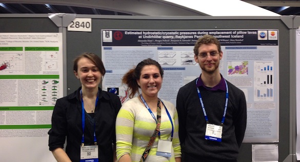
Above, Andy Nash (left, ’14) and Simon Doong (’15) are listening to Candy Thornton (right, ’14) talk about her project, which was inspired by Toure, a speaker at Wooster’s 2013 Forum Series called “Facing Race”.  Candy investigated the intersection of public transportation, unemployment, and ethnicity in Los Angeles County (CA).

DSC_2631Owen Yeazell (left, ’14) listens as Kyle Burden (right, ’14) discusses his project on natural hazard mitigation: the spatial comparison between population centers and volcanic centers in California.

DSC_2633Scott Kugel’s (left, ’14) poster is directly related to his I.S. research and an offshoot of his experience this past summer as a member of a Keck Geology Consortium project.  Scott is intensely explaining his analysis of Connecticut River discharge during Hurricane Irene to Cameron Matesich (right, ’14).

DSC_2634Zach Sheehan’s (above, ’14) 2013 summer internship/experiences peaked his interest on food deserts and food accessibility issues in Ohio.  He translated that to a project that analyzed Ohio median household income (by tracts) fast food restaurants, number of grocery stores, and obesity.

I am very proud of all of the GIS work this semester.  Not only did the students do a wonderful job presenting at the GIS Poster Symposium today, but in recent weeks, they also learned to navigate data problems and mysterious software issues along the way!!

In case you are interested in the amazing variety of projects, here is a list of students in GEOL 220 and their individual projects:

  • Kyle Burden, “A Spatial Comparison between Volcanoes and Areas of High Population in California, USA”
  • Allison Chin, “Analysis of Groundwater Pollution in Wayne County, OH”
  • Simon Doong, “Comparison of Military, Education, and Health Spending Among Nations”
  • Coleman Fitch, “Marcellus Shale Formation: Drilling Permits Relationship to Shale Depth and Productivity”
  • Cassandra Greenbaum, “Potential Influences for Obesity in the United States”
  • Perry Grosch, “Analysis of Human Populations around areas that have been marked by the incidents on the International Nuclear Event Scale in the United States before 2007”
  • Nichole Gustafson, “Forest Fragmentation and Garlic Mustard Colonization”
  • Tricia Hall, “Analysis of Fluid Flow in the Late Cretaceous Sixmile Canyon Formation”
  • Allison Ham, “The Influence of County Income on the Increasing Rate of Living Cases of HIV/AIDS in the D.C. Area”
  • Alex Hiatt, “Mapping Potential Jokulhlaup Flow Paths at Eyjafjallajokull in Southern Iceland”
  • Scott Kugel, “Maximum Discharge of the Connecticut River and its Tributaries During Hurricane Irene, August 21-30, 2011
  • Cameron Matesich, “GIS Model of the Geochemical Analysis of the High-Silica and Low-Silica Basalt Flows from Miter Crater in Ice Springs Volcanic Field, Black Rock Desert, Utah”
  • Stephanie Megas, “Portfolio Model School District Spatial Distribution and Projected Implications for Communities Surrounding School Closings in Baltimore City Public Schools (BCPS) using GIS Model Building”
  • Oscar Mmari, “Analysis of the Location of Federal Land, Population Density, Income, Water, and Fracking Wells in Utica and Marcellus Formations of Ohio”
  • Andy Nash, “Holocene Glacier Fluctuations Mapped in Glacier Bay National Park and Preserve using Radiocarbon Dated Detrital Logs”
  • Brian Porrett, “Putting World-Systems Analysis in Context: An Exploration of the Core and Periphery Relationship in the Early Bronze Age Levant”
  • Zachariah Sheehan, “The Impact of Income and Type of Food Accessibility on Obesity”
  • Ashleigh Sims, “How Does Grave Style Change Over Time and Space in the Cemetery of Athienou, Cyprus?”
  • Wyatt Smith, “Voting as a Function of Boredom: A State-by-State Analysis”
  • Nathan Taitano, “Wildlife Habitat and Land Management”
  • Candice Thornton, “ACESS:  A Socioeconomic GIS Study on the Impact of Public Transportation on Employment”
  • Jim Torpy, “Political Power and Mineral Access in Ancient Cyprus”
  • Henry Waldron, “ASU Downtown:  How a New Campus Affects Urban Property Values”
  • Owen Yeazell, “Impact of Roman Cities on Later English Populations”


Posted in Uncategorized | Tagged , , | Leave a comment